
Phospho-JNK1-T183/Y185 + JNK2-T183/Y185 + JNK3-T221/Y223 Rabbit mAb, Unconjugated
Catalog Number:
ABB-AP1337
- Images (8)

| Article Name: | Phospho-JNK1-T183/Y185 + JNK2-T183/Y185 + JNK3-T221/Y223 Rabbit mAb, Unconjugated |
| Biozol Catalog Number: | ABB-AP1337 |
| Supplier Catalog Number: | AP1337 |
| Alternative Catalog Number: | ABB-AP1337-100UL,ABB-AP1337-500UL,ABB-AP1337-1000UL,ABB-AP1337-20UL |
| Manufacturer: | ABclonal |
| Host: | Rabbit |
| Category: | Antikörper |
| Application: | ELISA, IF, IHC-P, WB |
| Species Reactivity: | Human |
| Immunogen: | Synthetic peptide. This information is considered to be commercially sensitive. |
| Conjugation: | Unconjugated |
| Alternative Names: | JNK1/2/3, SAPK, Phospho-JNK1-T183/Y185 + JNK2-T183/Y185 + JNK3-T221/Y223 |
| The protein encoded by this gene is a member of the MAP kinase family. MAP kinases act as an integration point for multiple biochemical signals, and are involved in a wide variety of cellular processes such as proliferation, differentiation, transcription regulation and development. This kinase is activated by various cell stimuli, and targets specific transcription factors, and thus mediates immediate-early gene expression in response to cell stimuli. The activation of this kinase by tumor-necrosis factor alpha (TNF-alpha) is found to be required for TNF-alpha induced apoptosis. This kinase is also involved in UV radiation induced apoptosis, which is thought to be related to cytochrom c-mediated cell death pathway. Studies of the mouse counterpart of this gene suggested that this kinase play a key role in T cell proliferation, apoptosis and differentiation. Several alternatively spliced transcript variants encoding distinct isoforms have been reported. [provided by RefSeq, Apr 2016] |
| Application Dilute: | WB,1:1000 - 1:5000|IF-P,1:50 - 1:200|IHC-P,1:50 - 1:200|ELISA,Recommended starting concentration is 1 µg/mL. Please optimize the concentration based on your specific assay requirements. |
| Application Notes: | Cross-Reactivity: Human,Mouse,Rat. ResearchArea: Protein phosphorylation,Protein phosphorylation,Protein phosphorylation. Shipping: Ice Bag |
|
|
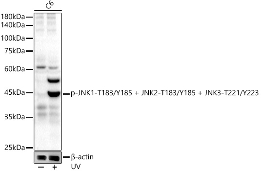
Western blot analysis of various lysates, using Phospho-JNK1-T183/Y185 + JNK2-T183/Y185 + JNK3-T221/Y223 Rabbit mAb (AP1337) at1:2000 dilution. C6 cells were treated with UV at room temperature for 15-30 minutes. Secondary antibody: HRP-conjugated Goat anti-Rabbit IgG (H+L) (AS014) at 1:10000 dilution. Lysates/proteins: 25µg per lane. Blocking buffer: 3% nonfat dry milk in TBST. Detection: ECL Basic Kit (RM00020). Exposure time: 30s. |
|
|
Immunohistochemistry analysis of paraffin-embedded Rat brain tissue using Phospho-JNK1-T183/Y185 + JNK2-T183/Y185 + JNK3-T221/Y223 Rabbit mAb (AP1337) at a dilution of 1:200 (40x lens). High pressure antigen retrieval performed with 0.01M Citrate buffer (pH 6.0) prior to IHC staining. |
|
|
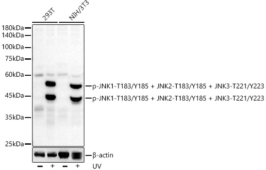
Western blot analysis of various lysates, using Phospho-JNK1-T183/Y185 + JNK2-T183/Y185 + JNK3-T221/Y223 Rabbit mAb (AP1337) at1:2000 dilution. 293T and NIH/3T3 cells were treated with UV at room temperature for 15-30 minutes. Secondary antibody: HRP-conjugated Goat anti-Rabbit IgG (H+L) (AS014) at 1:10000 dilution. Lysates/proteins: 25µg per lane. Blocking buffer: 3% nonfat dry milk in TBST. Detection: ECL Basic Kit (RM00020). Exposure time: 30s. |
|
|
Immunohistochemistry analysis of paraffin-embedded Mouse brain tissue using Phospho-JNK1-T183/Y185 + JNK2-T183/Y185 + JNK3-T221/Y223 Rabbit mAb (AP1337) at a dilution of 1:200 (40x lens). High pressure antigen retrieval performed with 0.01M Citrate buffer (pH 6.0) prior to IHC staining. |
|
|
Immunohistochemistry analysis of paraffin-embedded Mouse kidney tissue using Phospho-JNK1-T183/Y185 + JNK2-T183/Y185 + JNK3-T221/Y223 Rabbit mAb (AP1337) at a dilution of 1:200 (40x lens). High pressure antigen retrieval performed with 0.01M Citrate buffer (pH 6.0) prior to IHC staining. |
|
|
Immunohistochemistry analysis of paraffin-embedded Rat colon tissue using Phospho-JNK1-T183/Y185 + JNK2-T183/Y185 + JNK3-T221/Y223 Rabbit mAb (AP1337) at a dilution of 1:200 (40x lens). High pressure antigen retrieval performed with 0.01M Citrate buffer (pH 6.0) prior to IHC staining. |
|
|
Confocal imaging of paraffin-embedded Mouse brain tissue using Phospho-JNK1-T183/Y185 + JNK2-T183/Y185 + JNK3-T221/Y223 Rabbit mAb (AP1337, dilution 1:200) followed by a further incubation with Cy3 Goat Anti-Rabbit IgG (H+L) (AS007, dilution 1:500) (Red). DAPI was used for nuclear staining (Blue). Microwave antigen retrieval performed with 0.01M Citrate Buffer (pH 6.0) prior to IF staining. Objective: 40x. |
|
|
Confocal imaging of paraffin-embedded Rat brain tissue using Phospho-JNK1-T183/Y185 + JNK2-T183/Y185 + JNK3-T221/Y223 Rabbit mAb (AP1337, dilution 1:200) followed by a further incubation with Cy3 Goat Anti-Rabbit IgG (H+L) (AS007, dilution 1:500) (Red). DAPI was used for nuclear staining (Blue). Microwave antigen retrieval performed with 0.01M Citrate Buffer (pH 6.0) prior to IF staining. Objective: 40x. |
Product Guarantee and Expert Support
