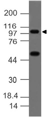
Fig:1- Expression analysis of A20. Anti-A20 antibody (Clone: ABM1G20) was tested at 4 µg/ml on THP-1 Lysate.
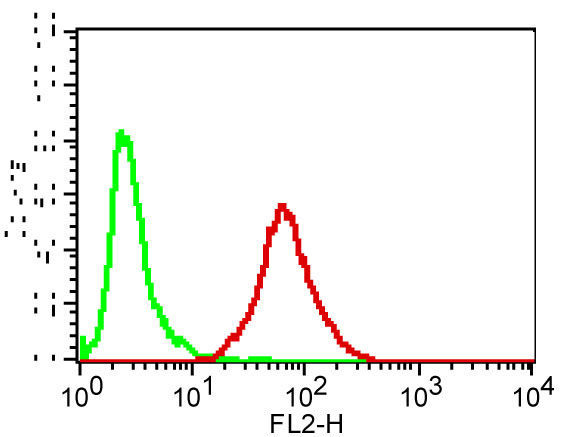
Fig-2: Cell surface FLOW analysis of A20 (Clone : ABM1G20) on HepG2 cells using 0.5 µg/10 6 cells of antibody. Goat anti-mouse PE conjugate was used as secondary antibody. Green represents isotope control (ABEOMICS), red represents anti-A20 antibody.
* VAT and and shipping costs not included. Errors and price changes excepted