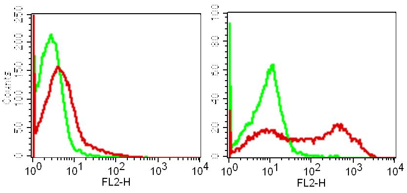
Figure-1: Cell surface flowcytometry analysis of human TIM-3 in PHA stimulated PBMC (Lymphocytes) using 0.5 µg/10 6 Cells of Anti-TIM-3 (Clone: ABM31A8) in Right and unstimulated PBMC is shown in Left. Green represent isotype control and Red represent Anti-TIM-3 antibody (ABEOMICS 10-4076). Goat antimouse PE conjugated was used as the secondary antibody.
* VAT and and shipping costs not included. Errors and price changes excepted