Monoclonal Antibody to Ikke/Ikki/TBK1 (Clone: ABM13C7)
Biozol Catalog Number:
ABI-10-6015-25
Supplier Catalog Number:
10-6015-25
Alternative Catalog Number:
ABI-10-6015-25-25UG
Manufacturer:
Abeomics
Host:
Mouse
Category:
Antikörper
Application:
FACS, IHC, WB
Species Reactivity:
Human, Mouse
Immunogen:
A partial length recombinant Ikke protein (amino acids 2-203) was used as the immunogen for this antibody.
Alternative Names:
IKBKE||IKKE||IKKI||KIAA0151
Ikke is a member of the IKK (IKappaB kinase) family that has been identified as an oncogenic protein and found to be up-regulated in breast cancer, ovarian cancer and prostate cancer. The Ikke protein has 33% and 31% identity at the amino acid level with Ikka and Ikkb, respectively and is primarily activated by interferon and mediates interferon signaling. The activated Ikke stimulates the transcription factor interferon regulatory factors 3 and phosphorylates signal transducer and activator of transcription. Ikke also shares some function with Ikka and Ikkb to activate NF- KappaB pathway by phosphorylation and degradation of IKappaBalpha. However, Ikke mainly mediates NF-KappaB activation induced by interferon, phorbol 12-myristate 13-acetate or the T-cell receptor, but not by tumor necrosis factor and interleukin 1, which activate Ikka and Ikkb. Ikke is predominantly expressed and active in peripheral blood leukocytes, pancreas, thymus, and spleen and contributes to IRF as well as NF-KappaB activation.
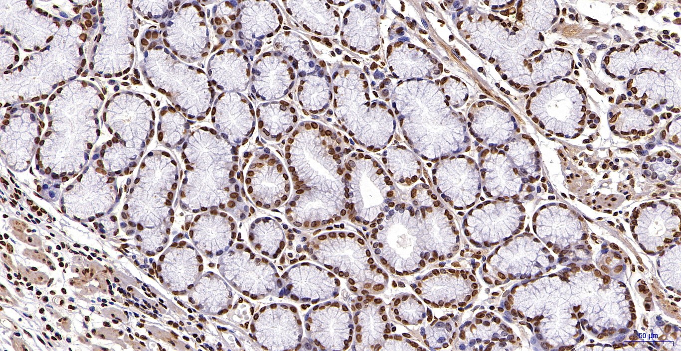
Western blot analysis: 2-4 µg/ml, Immunohistochemical analysis: 10-20 µg/ml
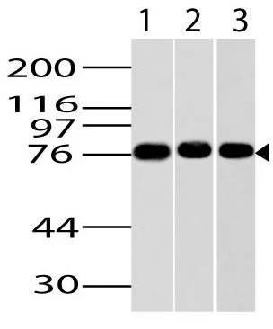
Fig-1: Western blot analysis of IKKe. Anti-Ikke antibody (Clone: ABM13C7) was tested at 2 µg/ml on Jurkat, NIH 3T3 and HepG2 lysates.
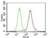
Fig-3: Intracellular staining of Jurkat cells. Green: Isotope control, Red: Anti-Ikke (10-6015) antibody. 0.5 ug of antibody was used. Goat anti-mouse PE was used as secondary antibody.
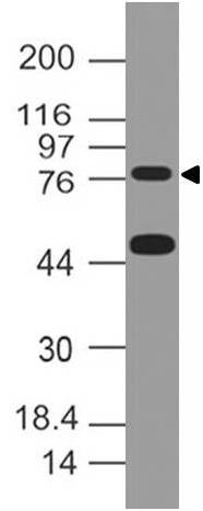
Fig-10: Western blot analysis of IKKe. Anti-Ikke antibody (Clone: ABM13C7) was tested at 1 µg/ml on m Liver lysate.
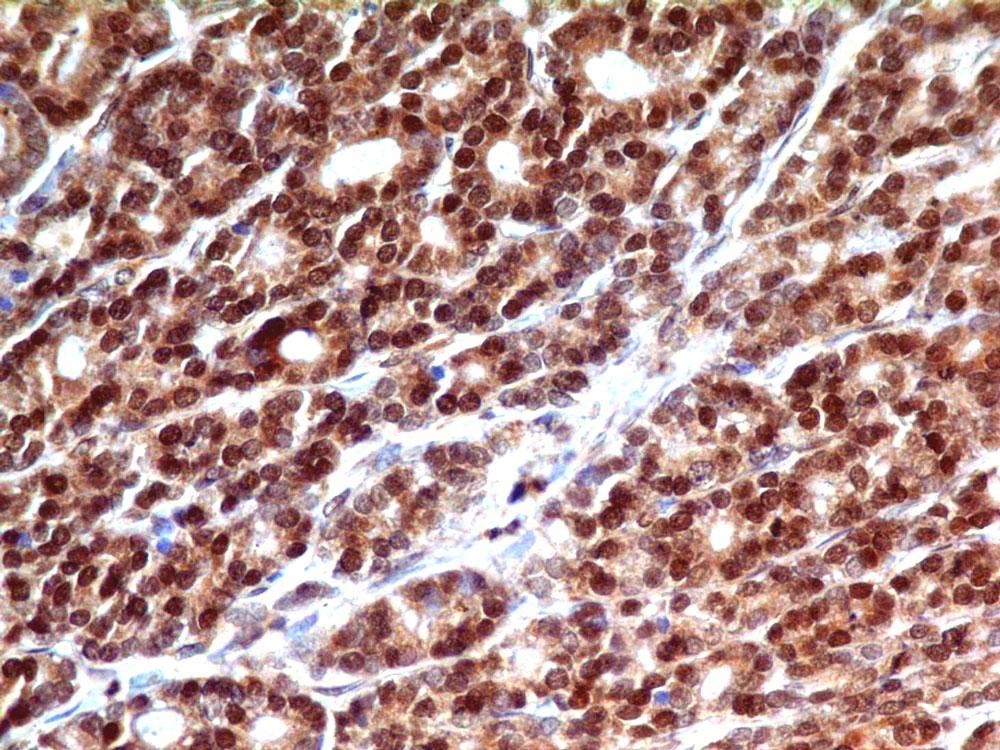
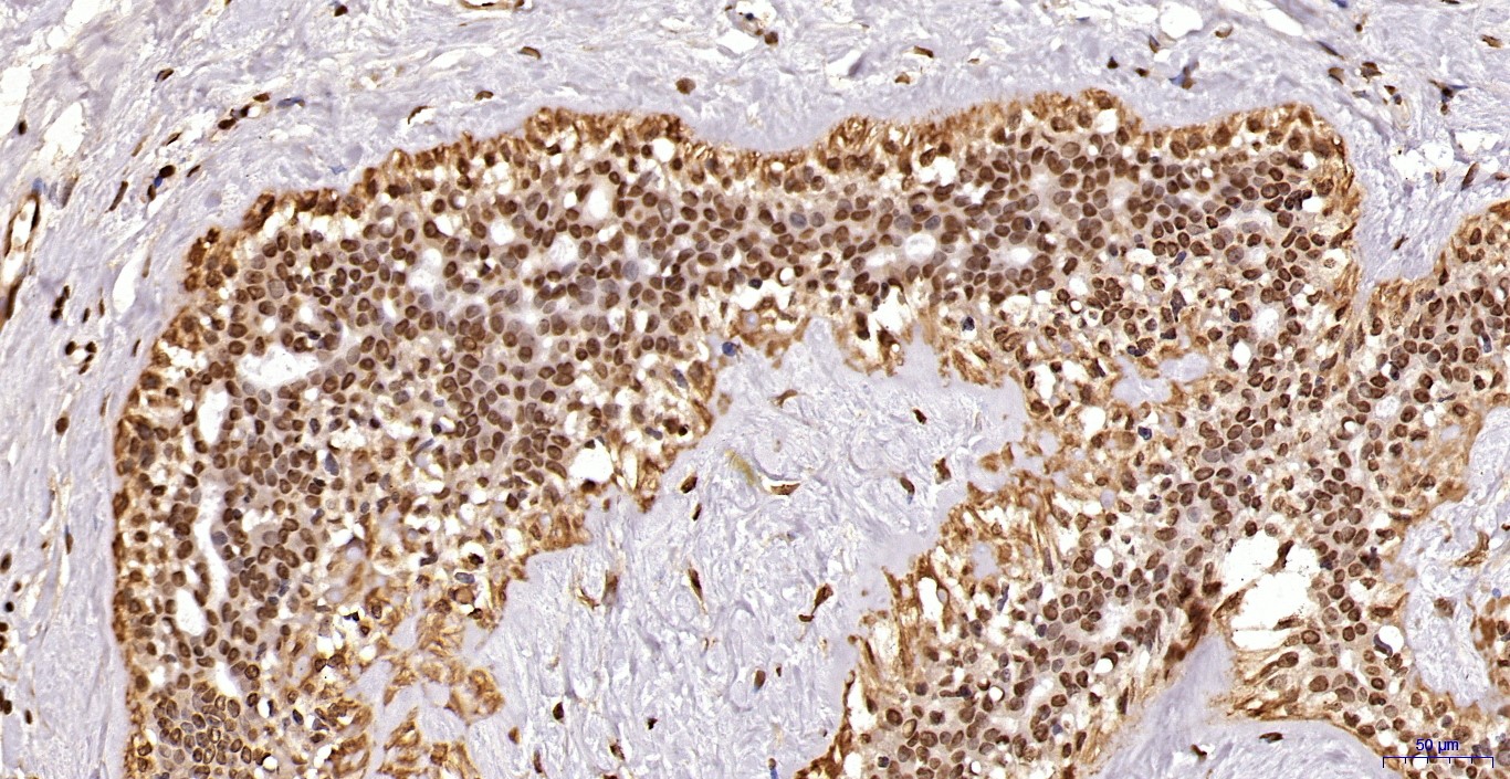
Fig-2: Immunohistochemical analysis of Ikke in Thyroid cancer tissue using Ikke antibody (Clone: ABM13C7) at 10 µg/ml.
Fig-4: Immunohistochemical analysis of Ikke in Breast cancer tissue using Ikke antibody (Clone: ABM13C7).
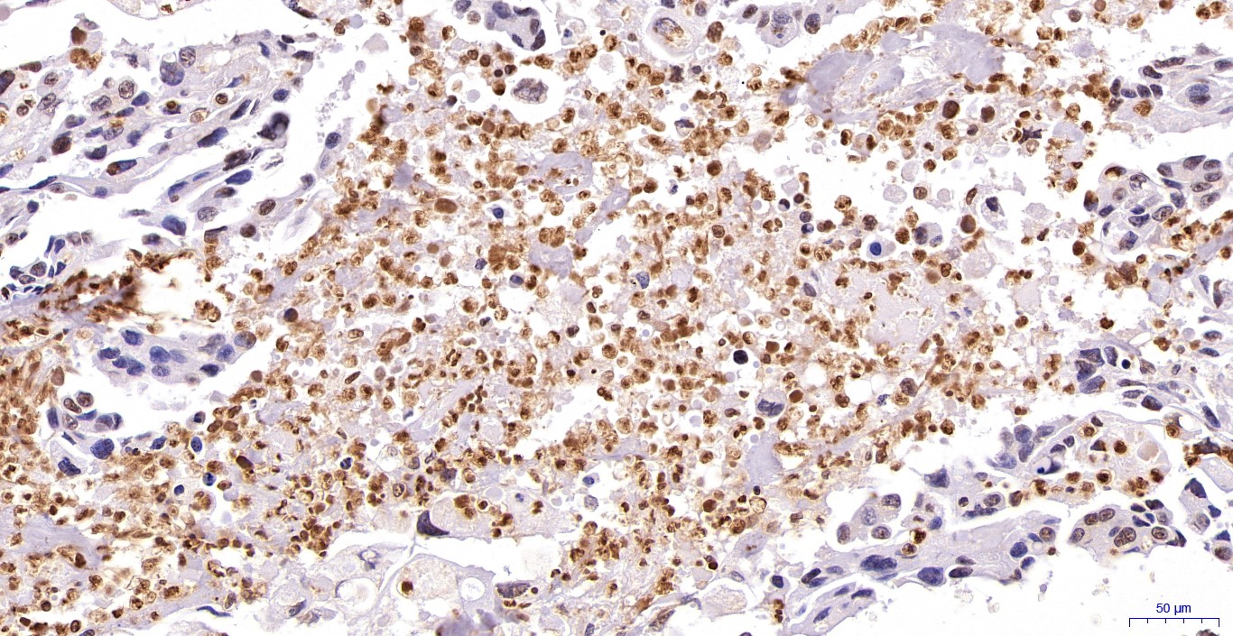
Fig-5: Immunohistochemical analysis of Ikke in Lungs cancer tissue using Ikke antibody (Clone: ABM13C7).
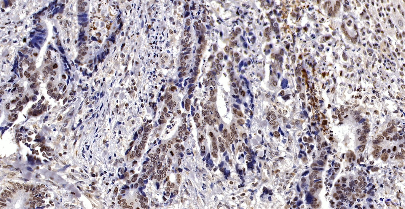
Fig-6: Immunohistochemical analysis of Ikke in Colon cancer tissue using Ikke antibody (Clone: ABM13C7).
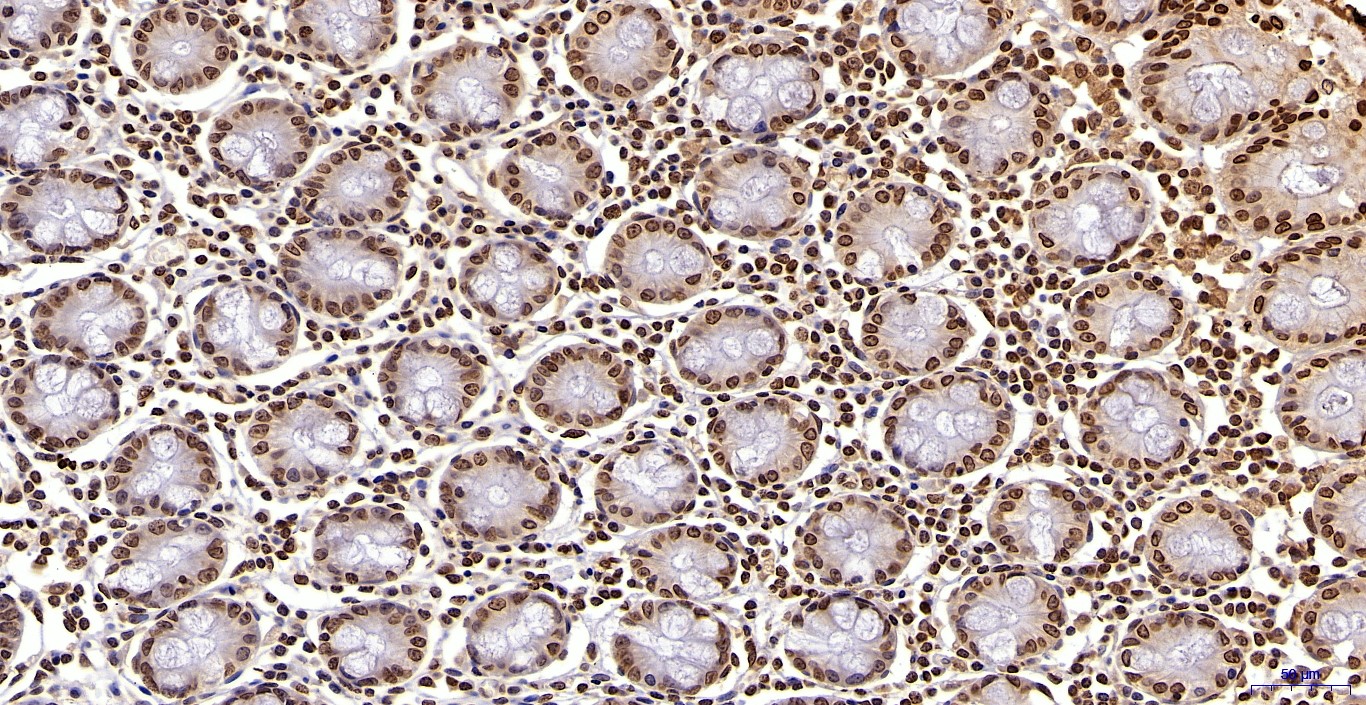
Fig-7: Immunohistochemical analysis of Ikke in Normal Colon tissue using Ikke antibody (Clone: ABM13C7).
Fig-8: Immunohistochemical analysis of Ikke in Normal Esophagus tissue using Ikke antibody (Clone: ABM13C7).
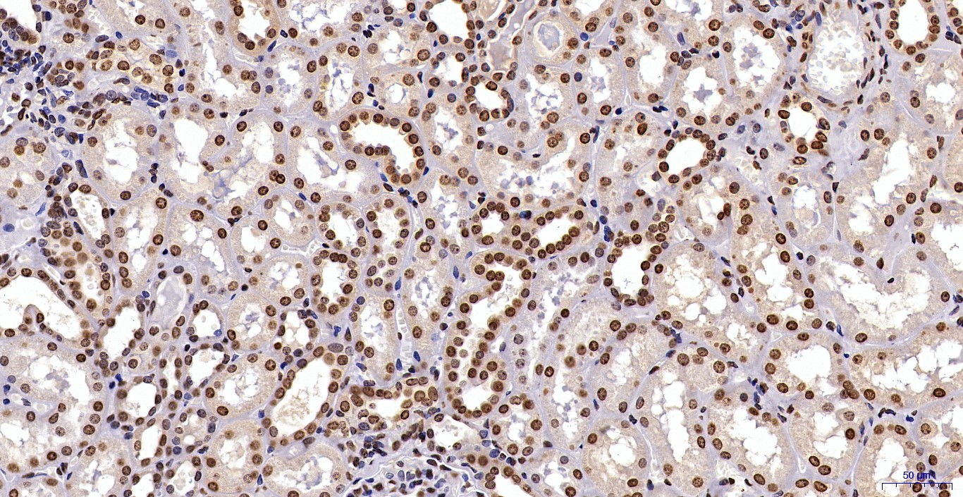
Fig-9: Immunohistochemical analysis of Ikke in Normal Kidney tissue using Ikke antibody (Clone: ABM13C7).
* VAT and and shipping costs not included. Errors and price changes excepted