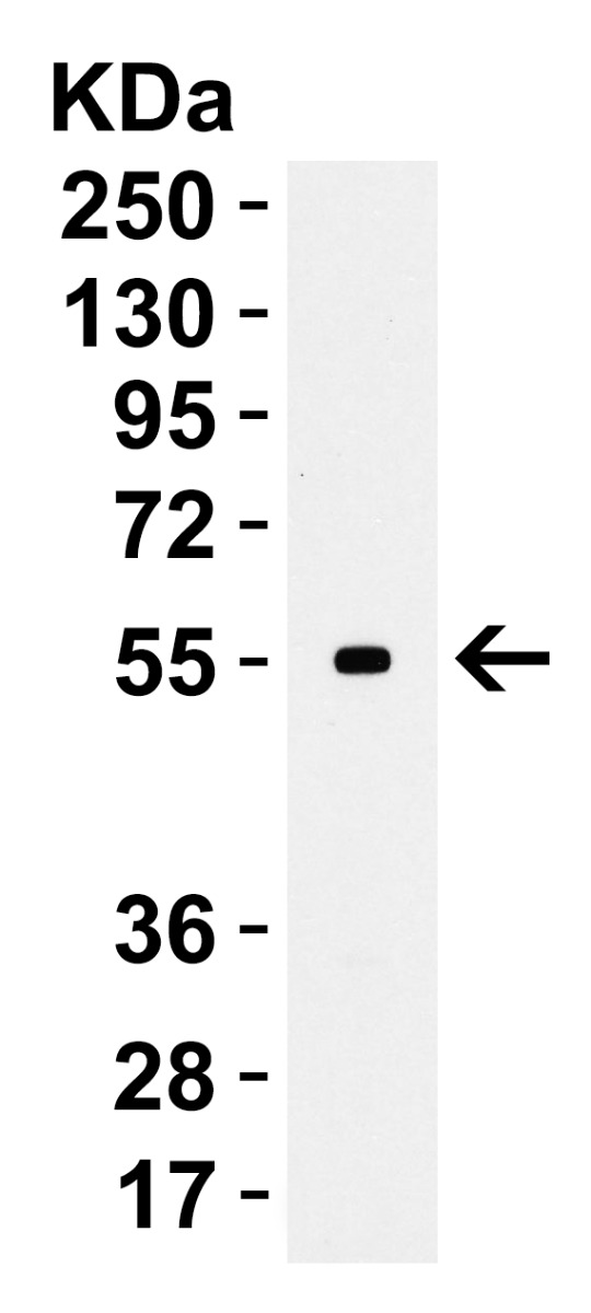
CD80 antibody was raised against a peptide corresponding to 17 amino acids near the amino terminus of human CD80. The immunogen is located within amino acids 60 - 110 of CD80.
CD80 Antibody is supplied in PBS containing 0.02% sodium azide.
Form:
Liquid
Application Dilute:
WB: 1-4 µg/mL, IHC-P/IF: 5-20 µg/mL. Antibody validated: Western Blot in human, mouse and rat samples, Immunofluorescence in the human, mouse and rat samples. All other applications and species not yet tested.
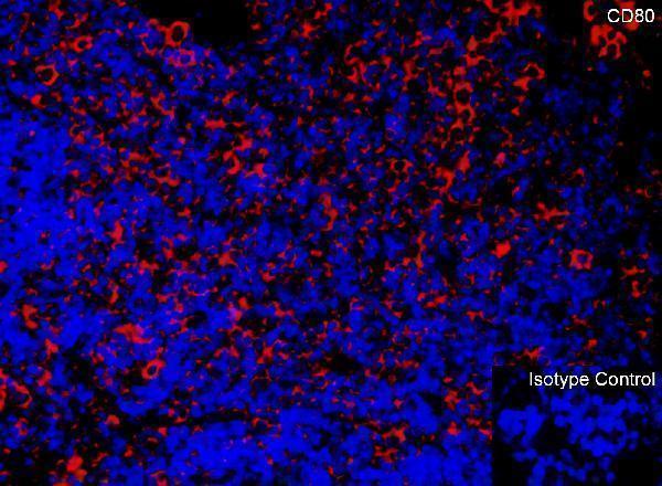
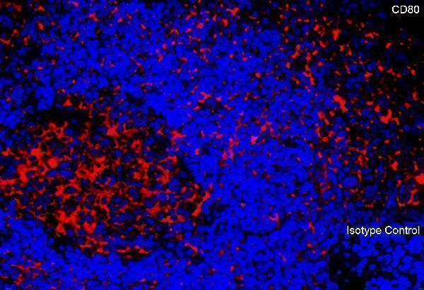
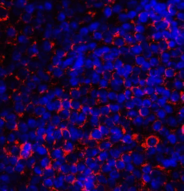
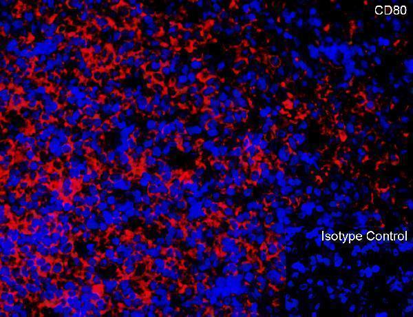
Immunofluorescence Validation of CD80 in Mouse SpleenImmunofluorescent analysis of 4% paraformaldehyde-fixed mouse spleen tissue labeling CD80 with A00196 at 20 µg/mL, followed by goat anti-rabbit IgG secondary antibody at 1/500 dilution (red) and DAPI staining (blue).
Immunofluorescence Validation of CD80 in Human TonsilImmunofluorescent analysis of 4% paraformaldehyde-fixed human tonsil tissue labeling CD80 with A00196 at 20 µg/mL, followed by goat anti-rabbit IgG secondary antibody at 1/500 dilution (red) and DAPI staining (blue).
Immunofluorescence Validation of CD80 in Human TonsilImmunofluorescent analysis of 4% paraformaldehyde-fixed human tonsil tissue labeling CD80 with A00196 at 20 µg/mL, followed by goat anti-rabbit IgG secondary antibody at 1/500 dilution (red) and DAPI staining (blue).
Immunofluorescence Validation of CD80 in Rat SpleenImmunofluorescent analysis of 4% paraformaldehyde-fixed rat spleen tissue labeling CD80 with A00196 at 20 µg/mL, followed by goat anti-rabbit IgG secondary antibody at 1/500 dilution (red) and DAPI staining (blue).
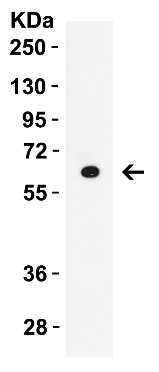
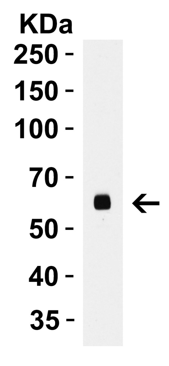
WB Validation in Human Raji CellsLoading: 15 µg of lysate Antibodies: CD80, A00196, 2 µ g/mL , 1 h incubation at RT in 5% NFDM/TBST. Secondary: Goat Anti-Rabbit IgG HRP conjugate at 1:10000 dilution.
WB Validation in Human ThymusLoading: 15 µg of lysate Antibodies: CD80, A00196, 2 µ g/mL , 1 h incubation at RT in 5% NFDM/TBST. Secondary: Goat Anti-Rabbit IgG HRP conjugate at 1:10000
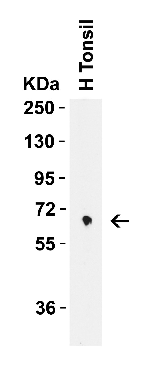
WB Validation in Human TonsilLoading: 15 µg of lysate Antibodies: CD80, A00196, 2 µg/mL , 1 h incubation at RT in 5% NFDM/TBST. Secondary: Goat Anti-Rabbit IgG HRP conjugate at 1:10000 dilution.
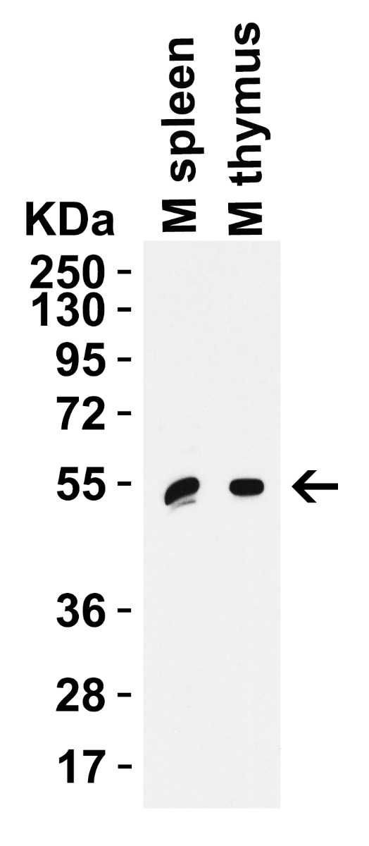
WB Validation in Mouse TissuesLoading: 15 µg of lysate Antibodies: CD80, A00196, 2 µg/mL , 1 h incubation at RT in 5% NFDM/TBST. Secondary: Goat Anti-Rabbit IgG HRP conjugate at 1:10000 dilution.
WB Validation in Rat ThymusLoading: 15 µg of lysate Antibodies: CD80, A00196, 2 µg/mL , 1 h incubation at RT in 5% NFDM/TBST. Secondary: Goat Anti-Rabbit IgG HRP conjugate at 1:10000 dilution.
* VAT and and shipping costs not included. Errors and price changes excepted