Boster Bio Anti-splicing factor 1/SF1 Antibody Picoband catalog A01009. Tested in ELISA, Flow Cytometry, IF, IHC, ICC, WB applications. This antibody reacts with Human, Mouse, Rat. The brand Picoband indicates this is a premium antibody that guarantees superior quality, high affinity, and strong signals with minimal background in Western blot applications. Only our best-performing antibodies are designated as Picoband, ensuring unmatched performance.
Clonality:
Polyclonal
Concentration:
Adding 0.2 ml of distilled water will yield a concentration of 500 µg/ml.
IHC analysis of splicing factor 1 using anti-splicing factor 1 antibody (A01009).splicing factor 1 was detected in paraffin-embedded section of mouse lung tissue. Heat mediated antigen retrieval was performed in citrate buffer (pH6&44, epitope retrieval solution) for 20 mins. The tissue section was blocked with 10% goat serum. The tissue section was then incubated with 1µg/ml rabbit anti-splicing factor 1 Antibody (A01009) overnight at 4C. Biotinylated goat anti-rabbit IgG was used as secondary antibody and incubated for 30 minutes at 37C. The tissue section was developed using Strepavidin-Biotin-Complex (SABC)(Catalog SA1022) with DAB as the chromogen.
IHC analysis of splicing factor 1 using anti-splicing factor 1 antibody (A01009).splicing factor 1 was detected in paraffin-embedded section of human placenta tissue. Heat mediated antigen retrieval was performed in citrate buffer (pH6&44, epitope retrieval solution) for 20 mins. The tissue section was blocked with 10% goat serum. The tissue section was then incubated with 1µg/ml rabbit anti-splicing factor 1 Antibody (A01009) overnight at 4C. Biotinylated goat anti-rabbit IgG was used as secondary antibody and incubated for 30 minutes at 37C. The tissue section was developed using Strepavidin-Biotin-Complex (SABC)(Catalog SA1022) with DAB as the chromogen.
IHC analysis of splicing factor 1 using anti-splicing factor 1 antibody (A01009).splicing factor 1 was detected in paraffin-embedded section of human rectal cancer tissue. Heat mediated antigen retrieval was performed in citrate buffer (pH6&44, epitope retrieval solution) for 20 mins. The tissue section was blocked with 10% goat serum. The tissue section was then incubated with 1µg/ml rabbit anti-splicing factor 1 Antibody (A01009) overnight at 4C. Biotinylated goat anti-rabbit IgG was used as secondary antibody and incubated for 30 minutes at 37C. The tissue section was developed using Strepavidin-Biotin-Complex (SABC)(Catalog SA1022) with DAB as the
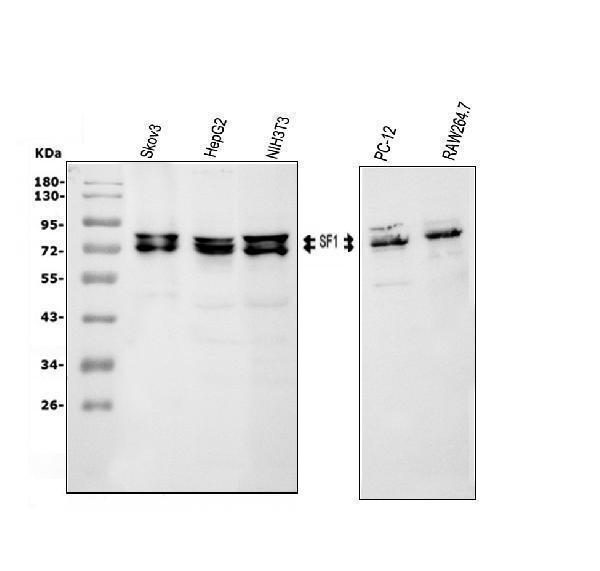
Western blot analysis of splicing factor 1 using anti-splicing factor 1 antibody (A01009). Electrophoresis was performed on a 5-20% SDS-PAGE gel at 70V (Stacking gel) / 90V (Resolving gel) for 2-3 hours. The sample well of each lane was loaded with 30 ug of sample under reducing conditions. Lane 1: human SK-OV-3 whole cell lysates, Lane 2: human HepG2 whole cell lysates, Lane 3: mouse NIH/3T3 whole cell lysates, Lane 4: rat PC-12 whole cell lysates, Lane 5: mouse RAW264.7 whole cell lysates. After electrophoresis, proteins were transferred to a nitrocellulose membrane at 150 mA for 50-90 minutes. Blocked the membrane with 5% non-fat milk/TBS for 1.5 hour at RT. The membrane was incubated with rabbit anti-splicing factor 1 antigen affinity purified polyclonal antibody (Catalog A01009) at 0.5 µg/mL overnight at 4C, then washed with TBS-0.1%Tween 3 times with 5 minutes each and probed with a goat anti-rabbit IgG-HRP secondary antibody at a dilution of 1:5000 for 1.5 hour at RT. The signal is developed using an Enhanced Chemiluminescent detection (ECL) kit (Catalog EK1002) with Tanon 5200 system. A specific band was detected for splicing factor 1 at approximately 68-80 kDa. The expected band size for splicing factor 1 is at 68 kDa.
IHC analysis of splicing factor 1 using anti-splicing factor 1 antibody (A01009).splicing factor 1 was detected in paraffin-embedded section of rat lung tissue. Heat mediated antigen retrieval was performed in citrate buffer (pH6&44, epitope retrieval solution) for 20 mins. The tissue section was blocked with 10% goat serum. The tissue section was then incubated with 1µg/ml rabbit anti-splicing factor 1 Antibody (A01009) overnight at 4C. Biotinylated goat anti-rabbit IgG was used as secondary antibody and incubated for 30 minutes at 37C. The tissue section was developed using Strepavidin-Biotin-Complex (SABC)(Catalog SA1022) with DAB as the chromogen.
* VAT and and shipping costs not included. Errors and price changes excepted