Boster Bio Anti-KDM6B/JMJD3 Picoband Antibody catalog A01309-1. Tested in ELISA, Flow Cytometry, WB applications. This antibody reacts with Human, Mouse. The brand Picoband indicates this is a premium antibody that guarantees superior quality, high affinity, and strong signals with minimal background in Western blot applications. Only our best-performing antibodies are designated as Picoband, ensuring unmatched performance.
Clonality:
Polyclonal
Concentration:
Adding 0.2 ml of distilled water will yield a concentration of 500 µg/ml.
Western blot, 0.25-0.5µg/ml, Human Flow Cytometry (Fixed), 1-3µg/1x106 cells, Human, Mouse ELISA, 0.1-0.5µg/ml, -
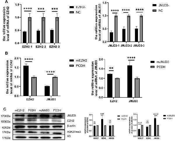
Expression levels of EZH2, JMJD3, H3K27me3 in spermatogonia. ( A ) qRT-PCR was used to detect the expression of EZH2 siRNA and JMJD3 siRNA in spermatogonia after interference. ( B ) measurements of EZH2 and JMJD3 mRNA levels in spermatogonia after overexpression. ( C ) The protein levels of EZH2, JMJD3 and H3K27me3 in spermatogonia were detected and statistically analyzed by Western blot. The membrane is lysed prior to hybridization with the antibody and the image has been cropped for a more aesthetically pleasing display. The full- length blots can be obtained from Additional file 2: Fig Index in PubMed under a CC BY license. PMID: 38424516
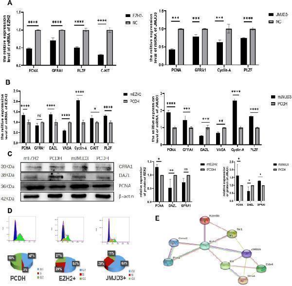
Effects of EZH2 interference or overexpression and JMJD3 interference or overexpression on self-renewal, proliferation and differentiation of spermatogonia. ( A ) The mRNA levels of PCNA, Cyclin-A, GFRA1, PLZF and C-KIT related to spermatogonia self-renewal and proliferation were changed after EZH2 and JMJD3 knockdown. ( B ) The expression of PCNA, cyclin-A, GFRA1, PLZF, C-KIT, DAZL and VASA was detected by qRT-PCR after EZH2 and JMJD3 overexpress
Flow Cytometry analysis of K562 cells using anti-KDM6B antibody (A01309-1). Overlay histogram showing K562 cells stained with A01309-1 (Blue line). To facilitate intracellular staining, cells were fixed with 4% paraformaldehyde and permeabilized with permeabilization buffer. The cells were blocked with 10% normal goat serum. And then incubated with rabbit anti-KDM6B Antibody (A01309-1, 1µg/1x106 cells) for 30 min at 20C. DyLight®,488 conjugated goat anti-rabbit IgG (BA1127, 5-10µg/1x106 cells) was used as secondary antibody for 30 minutes at 20C. Isotype control antibody (Green line) was rabbit IgG (1µg/1x106) used under the same conditions. Unlabelled sample without incubation with primary antibody and secondary antibody (Red line) was used as a blank control.
Flow Cytometry analysis of HEPA1-6 cells using anti-KDM6B antibody (A01309-1). Overlay histogram showing HEPA1-6 cells stained with A01309-1 (Blue line). To facilitate intracellular staining, cells were fixed with 4% paraformaldehyde and permeabilized with permeabilization buffer. The cells were blocked with 10% normal goat serum. And then incubated with rabbit anti-KDM6B Antibody (A01309-1, 1µg/1x106 cells) for 30 min at 20C. DyLight®,488 conjugated goat anti-rabbit IgG (BA1127, 5-10µg/1x106 cells) was used as secondary antibody for 30 minutes at 20C. Isotype control antibody (Green line) was rabbit IgG (1µg/1x106) used under the same conditions. Unlabelled sample without incubation with primary antibody and secondary antibody (Red line) was used as a blank control.
Western blot analysis of KDM6B using anti-KDM6B antibody (A01309-1). Electrophoresis was performed on a 5-20% SDS-PAGE gel at 70V (Stacking gel) / 90V (Resolving gel) for 2-3 hours. The sample well of each lane was loaded with 50ug of sample under reducing conditions. Lane 1: human K562 whole cell lysates, Lane 2: human A375 whole cell lysates, Lane 3: human HEK293 whole cell lysates, Lane 4: human A431 whole cell lysates. After Electrophoresis, proteins were transferred to a Nitrocellulose membrane at 150mA for 50-90 minutes. Blocked the membrane with 5% Non-fat Milk/ TBS for 1.5 hour at RT. The membrane was incubated with rabbit anti-KDM6B antigen affinity purified polyclonal antibody (Catalog A01309-1) at 0.5 µg/mL overnight at 4C, then washed with TBS-0.1%Tween 3 times with 5 minutes each and probed with a goat anti-rabbit IgG-HRP secondary antibody at a dilution of 1:5000 for 1.5 hour at RT. The signal is developed using an Enhanced Chemiluminescent detection (ECL) kit (Catalog EK1002) with Tanon 5200 system. A specific band was detected for KDM6B at approximately 177KD. The expected band size for KDM6B is at 177KD.
* VAT and and shipping costs not included. Errors and price changes excepted