E. coli-derived human FCGR2A recombinant protein (Position: Q34-M210).
Alternative Names:
CD32, CD32A, CDw32, Fc gamma RII a, Fc gamma RIIa, FCG2, FcGR, FCGR2, FCGR2A, FCGR2A1, FcRII a, IGFR2, IgG Fc receptor II a
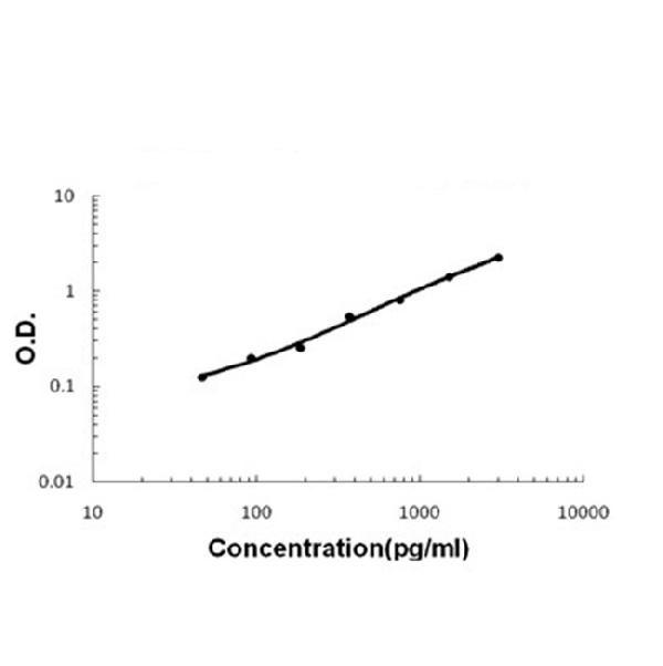
Boster Bio Anti-FCGR2A Antibody Picoband catalog A01450-1. Tested in ELISA, IHC, WB applications. This antibody reacts with Human, Mouse, Rat. The brand Picoband indicates this is a premium antibody that guarantees superior quality, high affinity, and strong signals with minimal background in Western blot applications. Only our best-performing antibodies are designated as Picoband, ensuring unmatched performance.
Clonality:
Polyclonal
Concentration:
Adding 0.2 ml of distilled water will yield a concentration of 500 µg/ml.
Each vial contains 4mg Trehalose, 0.9mg NaCl and 0.2mg Na2HPO4.
Purity:
Immunogen affinity purified.
Form:
Lyophilized
Target:
Low affinity immunoglobulin gamma Fc region receptor II-a
Application Dilute:
Western blot, 0.1-0.5µg/ml Immunohistochemistry (Paraffin-embedded Section), 0.5-1µg/ml ELISA (Cap), 1-5µg/ml
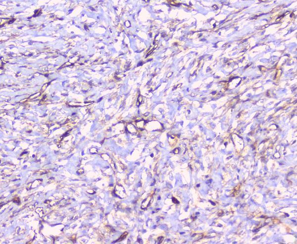
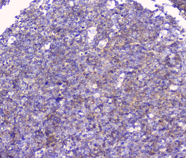
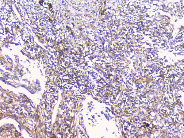
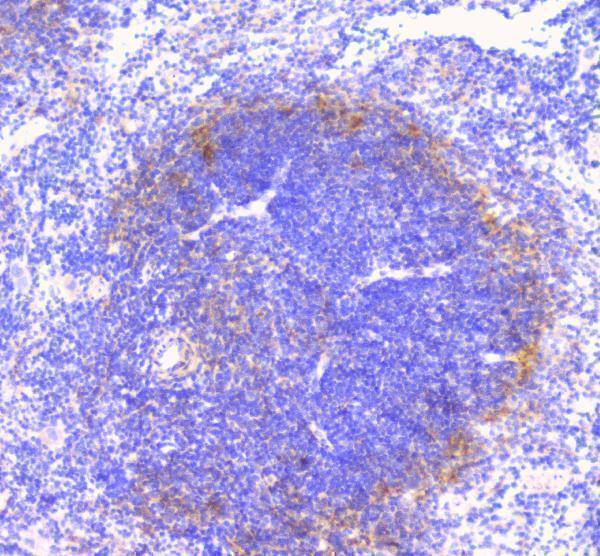
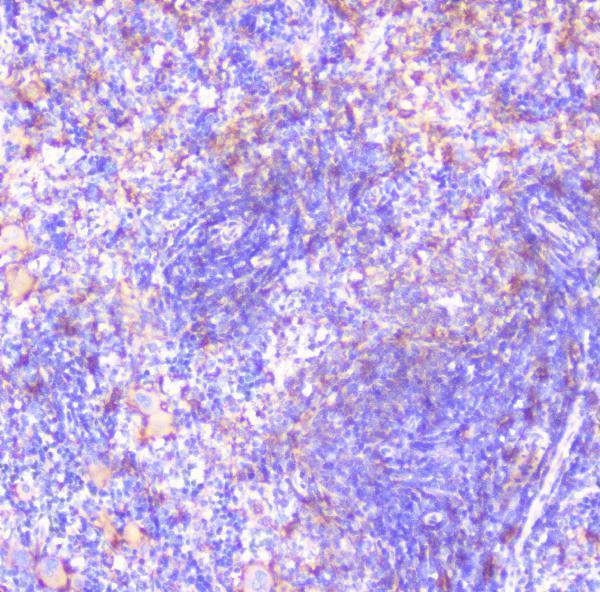
IHC analysis of FCGR2A using anti-FCGR2A antibody (A01450-1).FCGR2A was detected in paraffin-embedded section of human mammary cancer tissue. Heat mediated antigen retrieval was performed in citrate buffer (pH6&44, epitope retrieval solution) for 20 mins. The tissue section was blocked with 10% goat serum. The tissue section was then incubated with 1µg/ml rabbit anti-FCGR2A Antibody (A01450-1) overnight at 4C. Biotinylated goat anti-rabbit IgG was used as secondary antibody and incubated for 30 minutes at 37C. The tissue section was developed using Strepavidin-Biotin-Complex (SABC)(Catalog SA1022) with DAB as the chromogen.
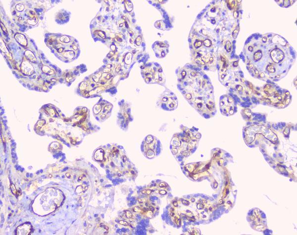
IHC analysis of FCGR2A using anti-FCGR2A antibody (A01450-1).FCGR2A was detected in paraffin-embedded section of human placenta tissue. Heat mediated antigen retrieval was performed in citrate buffer (pH6&44, epitope retrieval solution) for 20 mins. The tissue section was blocked with 10% goat serum. The tissue section was then incubated with 1µg/ml rabbit anti-FCGR2A Antibody (A01450-1) overnight at 4C. Biotinylated goat anti-rabbit IgG was used as secondary antibody and incubated for 30 minutes at 37C. The tissue section was developed using Strepavidin-Biotin-Complex (SABC)(Catalog SA1022) with DAB as the chromogen.
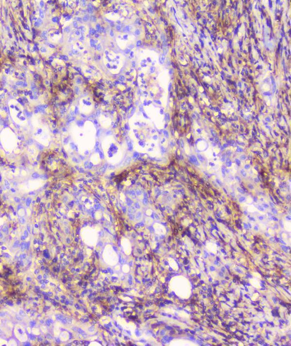
IHC analysis of FCGR2A using anti-FCGR2A antibody (A01450-1).FCGR2A was detected in paraffin-embedded section of human rectal cancer tissue. Heat m
IHC analysis of FCGR2A using anti-FCGR2A antibody (A01450-1).FCGR2A was detected in paraffin-embedded section of human tonsil tissue. Heat mediated antigen retrieval was performed in citrate buffer (pH6&44, epitope retrieval solution) for 20 mins. The tissue section was blocked with 10% goat serum. The tissue section was then incubated with 1µg/ml rabbit anti-FCGR2A Antibody (A01450-1) overnight at 4C. Biotinylated goat anti-rabbit IgG was used as secondary antibody and incubated for 30 minutes at 37C. The tissue section was developed using Strepavidin-Biotin-Complex (SABC)(Catalog SA1022) with DAB as the chromogen.
IHC analysis of FCGR2A using anti-FCGR2A antibody (A01450-1).FCGR2A was detected in paraffin-embedded section of human lung cancer tissue. Heat mediated antigen retrieval was performed in citrate buffer (pH6&44, epitope retrieval solution) for 20 mins. The tissue section was blocked with 10% goat serum. The tissue section was then incubated with 1µg/ml rabbit anti-FCGR2A Antibody (A01450-1) overnight at 4C. Biotinylated goat anti-rabbit IgG was used as secondary antibody and incubated for 30 minutes at 37C. The tissue section was developed using Strepavidin-Biotin-Complex (SABC)(Catalog SA1022) with DAB as the chromogen.
IHC analysis of FCGR2A using anti-FCGR2A antibody (A01450-1).FCGR2A was detected in paraffin-embedded section of mouse spleen tissue. Heat mediated antigen retrieval was performed in citrate buffer (pH6&44, epitope retrieval solution) for 20 mins. The tissue section was blocked with 10% goat serum. The tissue section was then incubated with 1µg/ml rabbit anti-FCGR2A Antibody (A01450-1) overnight at 4C. Biotinylated goat anti-rabbit IgG was used as secondary antibody and incubated for 30 minutes at 37C. The tissue section was developed using Strepavidin-Biotin-Complex (SABC)(Catalog SA1022) with DAB as the chromogen.
IHC analysis of FCGR2A using anti-FCGR2A antibody (A01450-1).FCGR2A was detected in paraffin-embedded section of rat spleen tissue. Heat mediated antigen retrieval was performed in citrate buffer (pH6&44, epitope retrieval solution) for 20 mins. The tissue section was blocked with 10% goat serum. The tissue section was then incubated with 1µg/ml rabbit anti-FCGR2A Antibody (A01450-1) overnight at 4C. Biotinylated goat anti-rabbit IgG was used as secondary antibody and incubated for 30 minutes at 37C. The tissue section was developed using Strepavidin-Biotin-Complex (SABC)(Catalog SA1022) with DAB as the chromogen.
* VAT and and shipping costs not included. Errors and price changes excepted