Borate Buffered Saline (BBS) pH 8.2 with 0.1% rAlbumin and 0.09% Sodium Azide
Form:
Liquid
Target:
JDP2
Application Dilute:
Application : Flow-Cyt , ApplicationDilutionRange : Fixed in 4% formaldehyde and permeabilized with 90% methanol. 1 µl per 1 x 10 6 cells. , Application : ICC , ApplicationDilutionRange : 1:40 to 1:100. Epitope retrieval with Tris-EDTA pH 9.0 is recommend
Application Notes:
Application Notes: Format: Whole IgG
Detection of mouse JDP2 (shaded) in CTLL-2 cells by flow cytometry. Antibody: Rabbit anti-JDP2 recombinant monoclonal antibody (orb1733245) or isotype control (unshaded). Secondary: DyLight 650-conjugated goat anti-rabbit IgG.
Detection of human JDP2 (shaded) in FaDu cells by flow cytometry. Antibody: Rabbit anti-JDP2 recombinant monoclonal antibody (orb1733245) or isotype control (unshaded). Secondary: DyLight 650-conjugated goat anti-rabbit IgG.
Detection of human JDP2 by immunocytochemistry. Sample: FFPE section of human FaDu cells. Antibody: Rabbit anti-JDP2 recombinant monoclonal antibody (orb1733245). Secondary: HRP-conjugated goat anti-rabbit IgG.
Detection of mouse JDP2 by immunocytochemistry. Sample: FFPE section of mouse C2C12 cells. Antibody: Rabbit anti-JDP2 recombinant monoclonal antibody (orb1733245). Secondary: HRP-conjugated goat anti-rabbit IgG.
Detection of human JDP2 by immunohistochemistry. Sample: FFPE section of human skin carcinoma. Antibody: Rabbit anti-JDP2 recombinant monoclonal antibody (orb1733245). Secondary: HRP-conjugated goat anti-rabbit IgG.
Detection of mouse JDP2 by immunohistochemistry. Sample: FFPE section of mouse RenCa tumor. Antibody: Rabbit anti-JDP2 recombinant monoclonal antibody (orb1733245). Secondary: HRP-conjugated goat anti-rabbit IgG.
Detection of human JDP2 by immunohistochemistry. Sample: FFPE section of human spleen. Antibody: Rabbit anti-JDP2 recombinant monoclonal antibody (orb1733245). Secondary: HRP-conjugated goat anti-rabbit IgG.
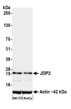
Detection of mouse JDP2 by western blot. Samples: Whole cell lysate (50 µg) from NIH 3T3 and RenCa cells prepared using NETN lysis buffer. Antibody: Rabbit anti-JDP2 recombinant monoclonal antibody (orb1733245) used at 1:1000. Secondary: HRP-conjugated goat anti-rabbit IgG. Detection: Chemiluminescence with an exposure time of 30 seconds. Lower Panel: Rabbit anti-Actin recombinant monoclonal antibody.
Detection of human JDP2 by western blot. Samples: Whole cell lysate (25 µg) from MCF-7, HeLa, HaCaT, and DU145 cells prepared using NETN lysis buffer. Antibody: Rabbit anti-JDP2 recombinant monoclonal antibody (orb1733245) used at 1:1000. Secondary: HRP-conjugated goat anti-rabbit IgG. Detection: Chemiluminescence with an exposure time of 3 minutes. Lower Panel: Rabbit anti-Actin recombinant monoclonal antibody.
* VAT and and shipping costs not included. Errors and price changes excepted