
HIST1H2AG (Ab-118) Antibody, Unconjugated, Rabbit, Polyclonal
Catalog Number:
CSB-PA010389OA118NFORHU
- Images (5)


| Article Name: | HIST1H2AG (Ab-118) Antibody, Unconjugated, Rabbit, Polyclonal |
| Biozol Catalog Number: | CSB-PA010389OA118NFORHU |
| Supplier Catalog Number: | CSB-PA010389OA118nforHU |
| Alternative Catalog Number: | CSB-PA010389OA118NFORHU-100UL, CSB-PA010389OA118NFORHU-50UL |
| Manufacturer: | Cusabio |
| Host: | Rabbit |
| Category: | Antikörper |
| Application: | ELISA, IF, IHC, IP, WB |
| Species Reactivity: | Human, Mouse |
| Conjugation: | Unconjugated |
| Alternative Names: | H2AC11 antibody, H2AFP antibody, HIST1H2AG, antibody, H2AC13 antibody, H2AFC antibody, HIST1H2AI, antibody, H2AC15 antibody, H2AFD antibody, HIST1H2AK, antibody, H2AC16 antibody, H2AFI antibody, HIST1H2AL, antibody, H2AC17 antibody, H2AFN antibody, HIST1H2AMHistone H2A type 1 antibody, H2A.1 antibody, Histone H2A/ptl antibody |
| Clonality: | Polyclonal |
| UniProt: | P0C0S8 |
| Buffer: | Preservative: 0.03% Proclin 300<br />Constituents: 50% Glycerol, 0.01M PBS, pH 7.4 |
| Purity: | Antigen Affinity Purified |
| Form: | Liquid |
| Target: | HIST1H2AG |
| Application Dilute: | Recommended dilution: WB:1:100-1:1000, IHC:1:10-1:100, IF:1:1-1:10, IP:1:200-1:2000 |
|
|
IHC image of CSB-PA010389OA118nforHU diluted at 1:20 and staining in paraffin-embedded human colon cancer performed on a Leica BondTM system. After dewaxing and hydration, antigen retrieval was mediated by high pressure in a citrate buffer (pH 6.0). Section was blocked with 10% normal goat serum 30min at RT. Then primary antibody (1% BSA) was incubated at 4°,C overnight. The primary is detected by a biotinylated secondary antibody and visualized using an HRP conjugated SP system. |
|
|
IHC image of CSB-PA010389OA118nforHU diluted at 1:20 and staining in paraffin-embedded human breast cancer performed on a Leica BondTM system. After dewaxing and hydration, antigen retrieval was mediated by high pressure in a citrate buffer (pH 6.0). Section was blocked with 10% normal goat serum 30min at RT. Then primary antibody (1% BSA) was incubated at 4°,C overnight. The primary is detected by a biotinylated secondary antibody and visualized using an HRP conjugated SP system. |
|
|
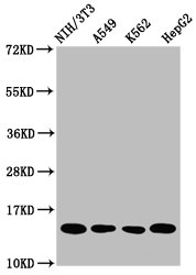
Western Blot Positive WB detected in: NIH/3T3 whole cell lysate, A549 whole cell lysate, K562 whole cell lysate, HepG2 whole cell lysate All lanes: HIST1H2AG antibody at 1µg/ml Secondary Goat polyclonal to rabbit IgG at 1/50000 dilution Predicted band size: 15 kDa Observed band size: 15 kDa |
|
|
Immunofluorescence staining of Hela cells with CSB-PA010389OA118nforHU at 1:5, counter-stained with DAPI. The cells were fixed in 4% formaldehyde, permeabilized using 0.2% Triton X-100 and blocked in 10% normal Goat Serum. The cells were then incubated with the antibody overnight at 4°,C. The secondary antibody was Alexa Fluor 488-congugated AffiniPure Goat Anti-Rabbit IgG(H+L). |
|
|
Immunoprecipitating HIST1H2AG in NIH/3T3 whole cell lysate Lane 1: Rabbit control IgG instead of CSB-PA010389OA118nforHU in NIH/3T3 whole cell lysate. For western blotting, a HRP-conjugated Protein G antibody was used as the secondary antibody (1/2000) Lane 2: CSB-PA010389OA118nforHU (5µg) + NIH/3T3 whole cell lysate (500µg) Lane 3: NIH/3T3 whole cell lysate (20µg) |
Product Guarantee and Expert Support
