The protein encoded by this gene belongs to the ser/thr protein kinase family. It is the catalytic subunit of the 5-prime-AMP-activated protein kinase (AMPK). AMPK is a cellular energy sensor conserved in all eukaryotic cells. The kinase activity of AMPK is activated by the stimuli that increase the cellular AMP/ATP ratio. AMPK regulates the activities of a number of key metabolic enzymes through phosphorylation. It protects cells from stresses that cause ATP depletion by switching off ATP-consuming biosynthetic pathways. Alternatively spliced transcript variants encoding distinct isoforms have been observed. [provided by RefSeq, Jul 2008],
Western Blot: 1/500 - 1/2000. Immunohistochemistry: 1/100 - 1/300. Immunofluorescence: 1/200 - 1/1000. ELISA: 1/10000. Not yet tested in other applications.
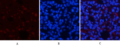
Immunofluorescence analysis of rat-lung tissue. 1,AMPKalpha1 Polyclonal Antibody(red) was diluted at 1:200(4C,overnight). 2, Cy3 labled Secondary antibody was diluted at 1:300(room temperature, 50min).3, Picture B: DAPI(blue) 10min. Picture A:Target. Picture B: DAPI. Picture C: merge of A+B
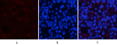
Immunofluorescence analysis of rat-lung tissue. 1,AMPKalpha1 Polyclonal Antibody(red) was diluted at 1:200(4C,overnight). 2, Cy3 labled Secondary antibody was diluted at 1:300(room temperature, 50min).3, Picture B: DAPI(blue) 10min. Picture A:Target. Picture B: DAPI. Picture C: merge of A+B
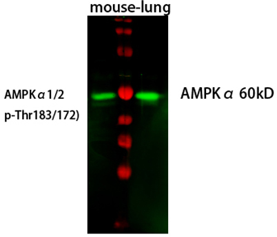
Western Blot analysis of mouse-lung cells using primary antibody diluted at 1:1000(4C overnight). Secondary antibody:Goat Anti-rabbit IgG IRDye 800( diluted at 1:5000, 25C, 1 hour). Cell lysate was extracted by Minute(TM) Plasma Membrane Protein Isolation and Cell Fractionation Kit(SM-005, Inventbiotech,MN,USA).
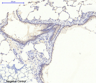
Immunohistochemical analysis of paraffin-embedded Rat-lung tissue. 1,AMPKalpha1 Polyclonal Antibody was diluted at 1:200(4C,overnight). 2, Sodium citrate pH 6.0 was used for antibody retrieval(>98C,20min). 3,Secondary antibody was diluted at 1:200(room tempeRature, 30min). Negative control was used by secondary antibody only.
* VAT and and shipping costs not included. Errors and price changes excepted