
Bcl-2 Antibody, Unconjugated, Rabbit, Polyclonal
Catalog Number:
PRS-3335
- Images (7)


| Article Name: | Bcl-2 Antibody, Unconjugated, Rabbit, Polyclonal |
| Biozol Catalog Number: | PRS-3335 |
| Supplier Catalog Number: | 3335 |
| Alternative Catalog Number: | PRS-3335-0.02,PRS-3335-0.1 |
| Manufacturer: | ProSci |
| Host: | Rabbit |
| Category: | Antikörper |
| Application: | ELISA, IF, IHC-P, WB |
| Species Reactivity: | Human, Mouse |
| Immunogen: | Anti-Bcl-2 antibody (3335) was raised against a peptide corresponding to 15 amino acids near the amino terminus of human Bcl-2. The immunogen is located within the first 50 amino acids of Bcl-2. |
| Conjugation: | Unconjugated |
| Alternative Names: | Bcl-2 Antibody: Bcl-2, PPP1R50, Apoptosis regulator Bcl-2 |
| Application Dilute: | Optimal dilutions for each application to be determined by the researcher. |
| Application Notes: | WB: 1-2µg/mL, IHC-P: 2 µg/mL, IF: 10 µg/mL.Antibody validated: Western Blot in human and mouse samples, Immunohistochemistry in human and mouse samples , Immunofluorescence in human and mouse samples. All other applications and species not yet tested. |
|
|
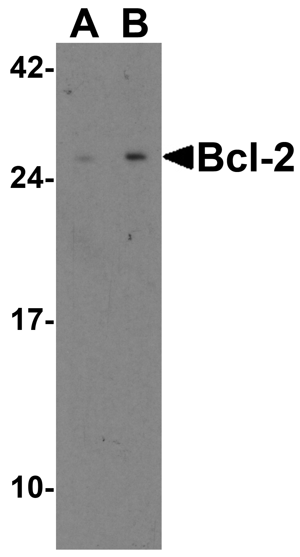
Figure 1 Western Blot Validation in Human and Mouse Cell Lines Loading: 15 &956,g of lysates per lane.Antibodies: Bcl-2 3335, (2 &956,g/mL), 1h incubation at RT in 5% NFDM/TBST.Secondary: Goat anti-rabbit IgG HRP conjugate at 1:10000 dilution.Lane A: Human Daudi cellsLane B: Mouse A-20 cells |
|
|
Figure 5 Immunofluorescence Validation of Bcl-2 in Human Kidney CellsImmunofluorescent analysis of 4% paraformaldehyde-fixed Human Kidney Cells labeling Bcl-2 with 3335 at 10 &956,g/mL, followed by goat anti-rabbit IgG secondary antibody at 1/500 dilution (red). |
|
|
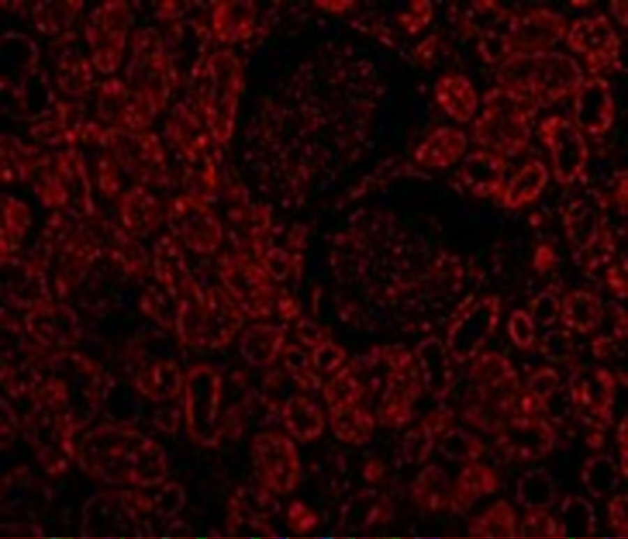
Figure 7 Immunofluorescence Validation of Bcl-2 in Mouse Brain TissueImmunofluorescent analysis of 4% paraformaldehyde-fixed Mouse Brain Tissue labeling Bcl-2 with 3335 at 20 &956,g/mL, followed by goat anti-rabbit IgG secondary antibody at 1/500 dilution (green). |
|
|
Figure 4 Immunohistochemistry Validation of Bcl-2 in Human Kidney Immunohistochemical analysis of paraffin-embedded Human Kidney using anti-Bcl-2 antibody (3335) at 2&956,g/ml. Tissue was fixed with formaldehyde and blocked with 10% serum for 1 h at RT, antigen retrieval was by heat mediation with a citrate buffer (pH6). Samples were incubated with primary antibody overnight at 4&730,C. A goat anti-rabbit IgG H&L (HRP) at 1/250 was used as secondary. Counter stained with Hematoxylin. |
|
|
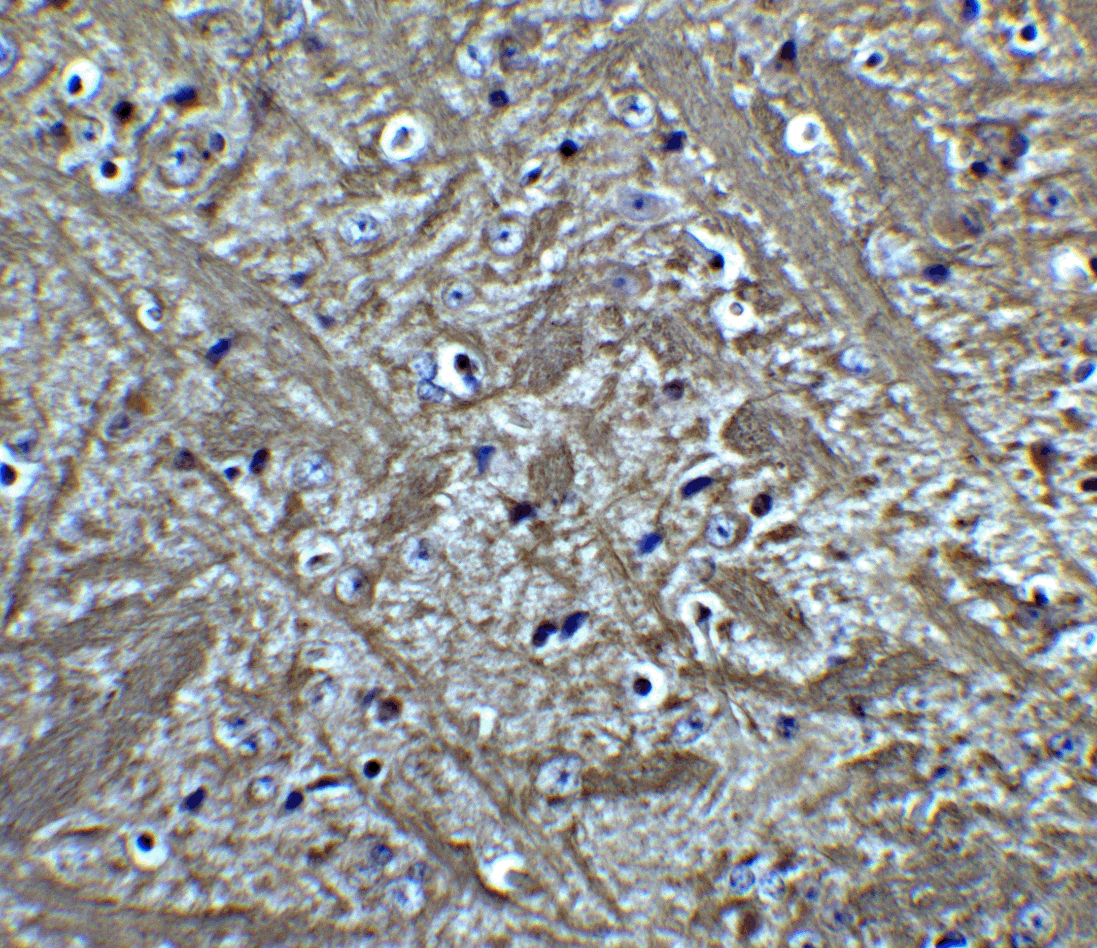
Figure 6 Immunohistochemistry Validation of Bcl-2 in Mouse Brain Tissue Immunohistochemical analysis of paraffin-embedded Mouse Brain Tissue using anti-Bcl-2 antibody (3335) at 5 &956,g/ml. Tissue was fixed with formaldehyde and blocked with 10% serum for 1 h at RT, antigen retrieval was by heat mediation with a citrate buffer (pH6). Samples were incubated with primary antibody overnight at 4&730,C. A goat anti-rabbit IgG H&L (HRP) at 1/250 was used as secondary. Counter stained with Hematoxylin. |
|
|
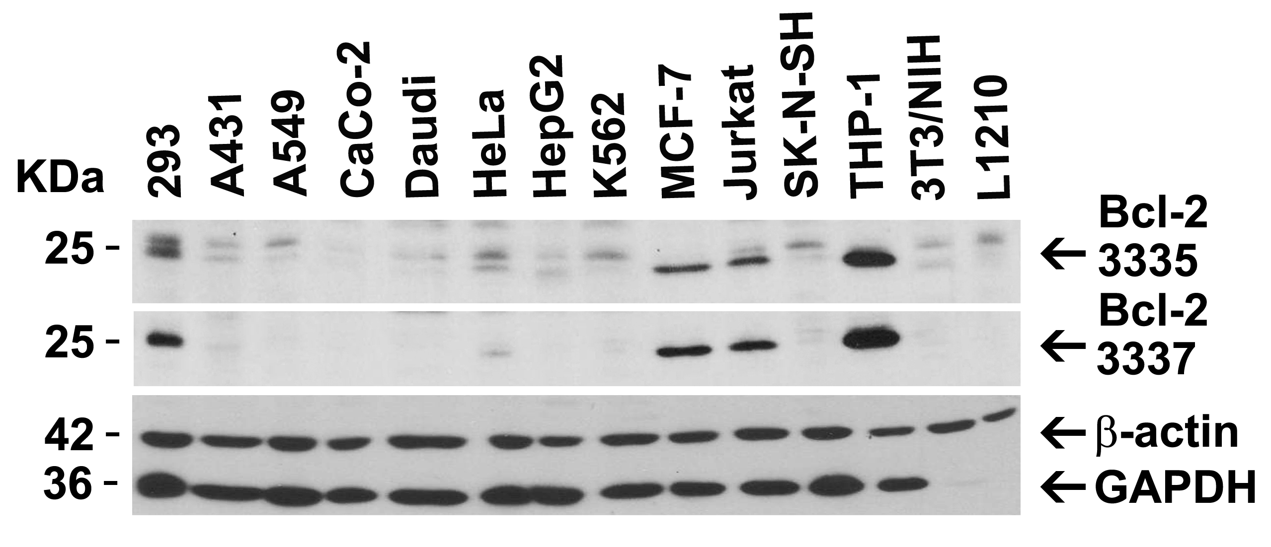
Figure 2 Independent Antibody Validation (IAV) via Protein Expression Profile in Cell LinesLoading: 15 &956,g of lysates per lane.Antibodies: Bcl-2 3335, (2 &956,g/mL), Bcl-2 3337, (2 &956,g/mL), beta-actin (1 &956,g/mL) and GAPDH (0.02 &956,g/mL), 1h incubation at RT in 5% NFDM/TBST.Secondary: Goat anti-rabbit IgG HRP conjugate at 1:10000 dilution. |
|
|
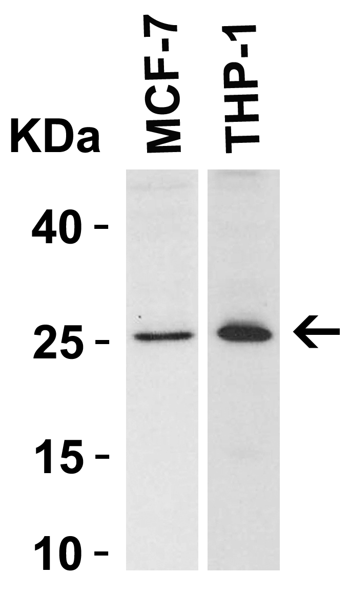
Figure 3 Western Blot Validation in Human Cell LinesLoading: 15 &956,g of lysates per lane.Antibodies: Bcl-2 3335, (2 &956,g/mL), 1h incubation at RT in 5% NFDM/TBST.Secondary: Goat anti-rabbit IgG HRP conjugate at 1:10000 dilution. |
Product Guarantee and Expert Support
