
IL-17 Antibody, Unconjugated, Rabbit, Polyclonal
Catalog Number:
PRS-4887
- Images (8)
| Article Name: | IL-17 Antibody, Unconjugated, Rabbit, Polyclonal |
| Biozol Catalog Number: | PRS-4887 |
| Supplier Catalog Number: | 4887 |
| Alternative Catalog Number: | PRS-4887-0.02,PRS-4887-0.1 |
| Manufacturer: | ProSci |
| Host: | Rabbit |
| Category: | Antikörper |
| Application: | ELISA, IF, IHC-P, WB |
| Species Reactivity: | Human, Mouse |
| Immunogen: | Anti-IL-17 antibody (4887) was raised against a peptide corresponding to 16 amino acids near the center of mature human IL-17. The immunogen is located within amino acids 50-100 of IL-17. |
| Conjugation: | Unconjugated |
| Alternative Names: | IL-17 Antibody: IL17, CTLA8, IL-17, IL-17A, IL17, Interleukin-17A, Cytotoxic T-lymphocyte-associated antigen 8 |
| Application Dilute: | Optimal dilutions for each application to be determined by the researcher. |
| Application Notes: | WB: 0.125-2 µg/mL, IF: 5-20 µg/mL, IHC: 1-5 µg/mL .Antibody validated: Western Blot in human samples, Immunofluorescence in mouse samples, Immunohistochemistry in human and mouse samples. All other applications and species not yet tested. |
|
|
Figure 5 Immunofluorescence Validation of IL-17 in Mouse A-20 CellsImmunofluorescent analysis of 4% paraformaldehyde-fixed mouse A-20 Cells labeling IL-17 with 4887 at 5 &956,g/mL, followed by goat anti-rabbit IgG secondary antibody at 1/500 dilution (green) and DAPI staining (blue). |
|
|
Figure 6 Immunofluorescence Validation of IL-17 in Mouse Thymus TissueImmunofluorescent analysis of 4% paraformaldehyde-fixed mouse thymus tissue labeling IL-17 with 4887 at 20 &956,g/mL, followed by goat anti-rabbit IgG secondary antibody at 1/500 dilution (red). |
|
|
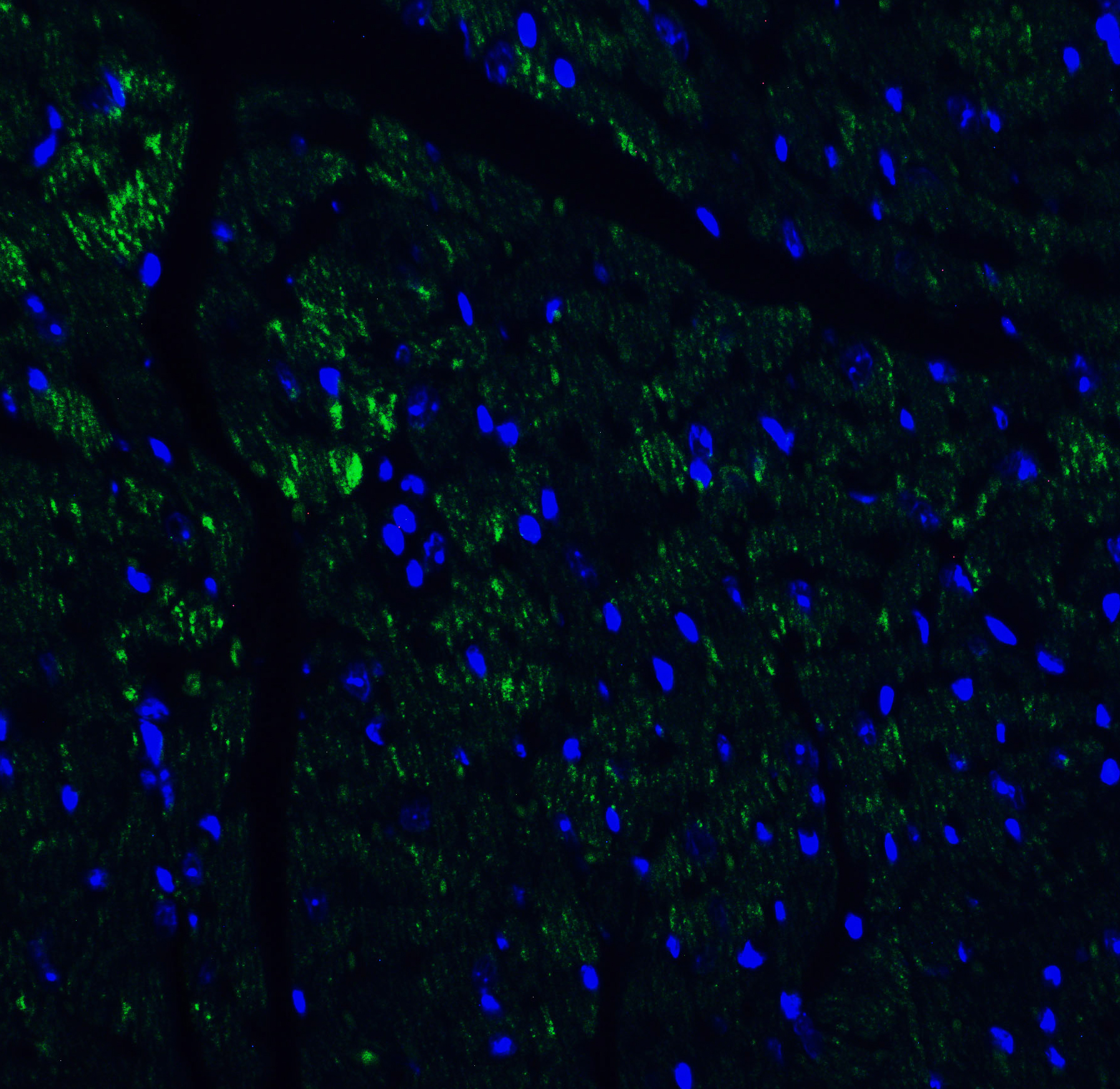
Figure 4 Immunofluorescence Validation of IL-17 in Mouse Heart TissueImmunofluorescent analysis of 4% paraformaldehyde-fixed mouse heart issue labeling IL-17 with 4887 at 10 &956,g/mL, followed by goat anti-rabbit IgG secondary antibody at 1/500 dilution (green) and DAPI staining (blue). |
|
|
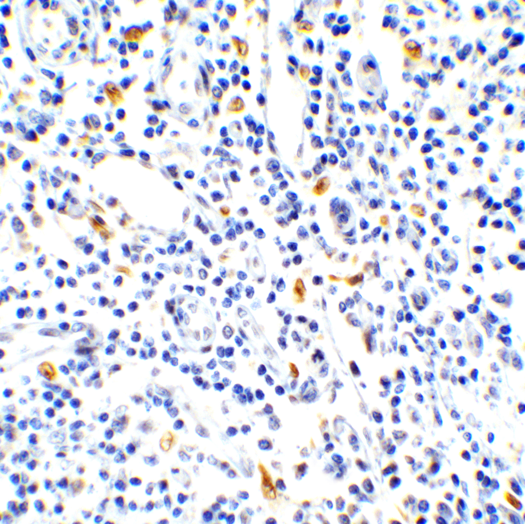
Figure 8 Immunohistochemistry Validation of IL-17 in Human Tonsil Tissue Immunohistochemical analysis of paraffin-embedded human tonsil tissue using anti-IL-17 antibody (4887) at 5 &956,g/mL. Tissue was fixed with formaldehyde and blocked with 10% serum for 1 h at RT, antigen retrieval was by heat mediation with a citrate buffer (pH6). Samples were incubated with primary antibody overnight at 4&730, C. A goat anti-rabbit IgG H&L (HRP) at 1/250 was used as secondary. Counter stained with Hematoxylin. |
|
|
Figure 7 Immunohistochemistry Validation of IL-17 in Mouse Colon Tissue Immunohistochemical analysis of paraffin-embedded mouse colon tissue using anti-IL-17 antibody (4887) at 2 &956,g/mL. Tissue was fixed with formaldehyde and blocked with 10% serum for 1 h at RT, antigen retrieval was by heat mediation with a citrate buffer (pH6). Samples were incubated with primary antibody overnight at 4&730, C. A goat anti-rabbit IgG H&L (HRP) at 1/250 was used as secondary. Counter stained with Hematoxylin. |
|
|
Figure 3 Immunohistochemistry Validation of IL-17 in Human Lymph Node Immunohistochemical analysis of paraffin-embedded human lymph node tissue using anti-IL-17 antibody (4887) at 1 &956,g/ml. Tissue was fixed with formaldehyde and blocked with 10% serum for 1 h at RT, antigen retrieval was by heat mediation with a citrate buffer (pH6). Samples were incubated with primary antibody overnight at 4&730,C. A goat anti-rabbit IgG H&L (HRP) at 1/250 was used as secondary. Counter stained with Hematoxylin. |
|
|
Figure 1 Western Blot Validation with Recombinant ProteinLoading: 30 ng of human IL-17 recombinant protein per lane.Antibodies: IL-17 4887, 1h incubation at RT in 5% NFDM/TBST.Secondary: Goat anti-rabbit IgG HRP conjugate at 1:10000 dilution.Lane 1: 0.125 &956,g/mLLane 2: 0.25 &956,g/mLLane 3: 0.5 &956,g/mL |
|
|
Figure 2 Western Blot Validation with Human Spleen Loading: 10 &956,g of lysates per lane.Antibodies: IL-17 4887, 1h incubation at RT in 5% NFDM/TBST.Secondary: Goat anti-rabbit IgG HRP conjugate at 1:10000 dilution.Exposure: 1 minLane 1: 1 &956,g/mL Lane 2: 2 &956,g/mL |
Product Guarantee and Expert Support
