
CD4 Antibody [9H5A8], IgG1, Unconjugated, Mouse, Monoclonal
Catalog Number:
PRS-PM-5201
- Images (8)


| Article Name: | CD4 Antibody [9H5A8], IgG1, Unconjugated, Mouse, Monoclonal |
| Biozol Catalog Number: | PRS-PM-5201 |
| Supplier Catalog Number: | PM-5201 |
| Alternative Catalog Number: | PRS-PM-5201-0.02,PRS-PM-5201-0.1 |
| Manufacturer: | ProSci |
| Host: | Mouse |
| Category: | Antikörper |
| Application: | ELISA, IHC-P, WB |
| Species Reactivity: | Human, Mouse, Rat |
| Immunogen: | Mouse monoclonal CD4 antibody was raised against a 193 amino acid recombinant protein from near the amino terminus of human CD4. |
| Conjugation: | Unconjugated |
| Alternative Names: | CD4 Antibody [9H5A8] : CD4mut |
| Clonality: | Monoclonal |
| Concentration: | 1 mg/mL |
| Clone Designation: | [9H5A8] |
| Molecular Weight: | Predicted: 51kDObserved: 51-57 kD (Post-modyfication: 2-4 N-linked glycosylations) |
| Isotype: | IgG1 |
| NCBI: | 920 |
| UniProt: | P01730 |
| Buffer: | CD4 Monoclonal Antibody is supplied in PBS containing 0.02% sodium azide. |
| Form: | Liquid |
| Application Dilute: | Optimal dilutions for each application to be determined by the researcher. |
| Application Notes: | WB: 0.5-4 µg/mL, IHC: 5 µg/mL.Antibody validated: Western Blot in human, mouse and rat samples, Immunohistochemistry in human samples. All other applications and species not yet tested. |
|
|
Figure 8 Immunohistochemistry Validation of CD4 in Human Thymus Tissue Immunohistochemical analysis of paraffin-embedded human thymus tissue using anti-CD4 antibody (PM-5201) at 5 &956,g/ml. Tissue was fixed with formaldehyde and blocked with 10% serum for 1 h at RT, antigen retrieval was by heat mediation with a citrate buffer (pH6). Samples were incubated with primary antibody overnight at 4&730,C. A goat anti-mouse IgG H&L (HRP) at 1/250 was used as secondary. Counter stained with Hematoxylin. |
|
|
Figure 1 Western Blot Validation with Human Recombinant ProteinLoading: 5ng (Lane 1), 25ng (Lane 2) and 100ng (Lane 3) of human CD4 recombinant protein.Antibodies: CD4 PM-5201 (1 &956,g/mL), 1h incubation at RT in 5% NFDM/TBST.Secondary: Goat anti-mouse IgG HRP conjugate at 1:5000 dilution.Observed at around 27kD. |
|
|
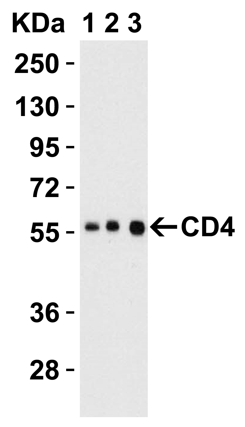
Figure 2 Western Blot Validation in Human Thymus Tissue LysateLoading: 15 &956,g of lysates per lane.Antibodies: CD4 PM-5201 (Lane 1: 0.5 &956,g/mL, Lane 2: 1 &956,g/mL and Lane 3: 2 &956,g/mL), 1h incubation at RT in 5% NFDM/TBST.Secondary: Goat anti-mouse IgG HRP conjugate at 1:5000 dilution. |
|
|
Figure 3 Western Blot Validation in Human Jurkat LysateLoading: 15 &956,g of lysates per lane.Antibodies: CD4 PM-5201 (Lane 1: 1 &956,g/mL, Lane 2: 2 &956,g/mL and Lane 3: 4 &956,g/mL), 1h incubation at RT in 5% NFDM/TBST.Secondary: Goat anti-mouse IgG HRP conjugate at 1:5000 dilution. |
|
|
Figure 4 Western Blot Validation in Human, Mouse and Rat Tissue LysateLoading: 15 &956,g of lysates per lane.Antibodies: CD4 PM-5201 (1 &956,g/mL), 1h incubation at RT in 5% NFDM/TBST.Secondary: Goat anti-mouse IgG HRP conjugate at 1:5000 dilution. |
|
|
Figure 5 Western Blot Validation in Human Cell Lines Loading: 15 &956,g of lysates per lane.Antibodies: CD4 PM-5201 (4 &956,g/mL), 1h incubation at RT in 5% NFDM/TBST.Secondary: Goat anti-mouse IgG HRP conjugate at 1:5000 dilution. |
|
|
Figure 6 Western Blot Validation in Mouse Spleen Tissue LysateLoading: 15 &956,g of lysates per lane.Antibodies: CD4 PM-5201 (Lane 1: 1 &956,g/mL, Lane 2: 2 &956,g/mL and Lane 3: 4 &956,g/mL), 1h incubation at RT in 5% NFDM/TBST.Secondary: Goat anti-mouse IgG HRP conjugate at 1:5000 dilution. |
|
|
Figure 7 Western Blot Validation in Rat Lung Tissue LysateLoading: 15 &956,g of lysates per lane.Antibodies: CD4 PM-5201 (Lane 1: 0.5 &956,g/mL, Lane 2: 1 &956,g/mL and Lane 3: 2 &956,g/mL), 1h incubation at RT in 5% NFDM/TBST.Secondary: Goat anti-mouse IgG HRP conjugate at 1:5000 dilution. |
Product Guarantee and Expert Support
