PD-L1 Antibody is supplied in PBS containing 0.02% sodium azide and 50% glycerol.
Form:
Liquid
Application Dilute:
Optimal dilutions for each application to be determined by the researcher.
Application Notes:
PD-L1 antibody can be used for detection of PD-L1 by Western blot at 0.5 - 1 µg/mL. Antibody can also be used for immunohistochemistry starting at 2 - 5 µg/mL. For immunofluorescence start at 5µg/mL.Antibody validated: Western Blot in human samples, Immunohistochemistry in human samples, Immunocytochemistry in human samples and Immunofluorescence in human samples. All other applications and species not yet tested.
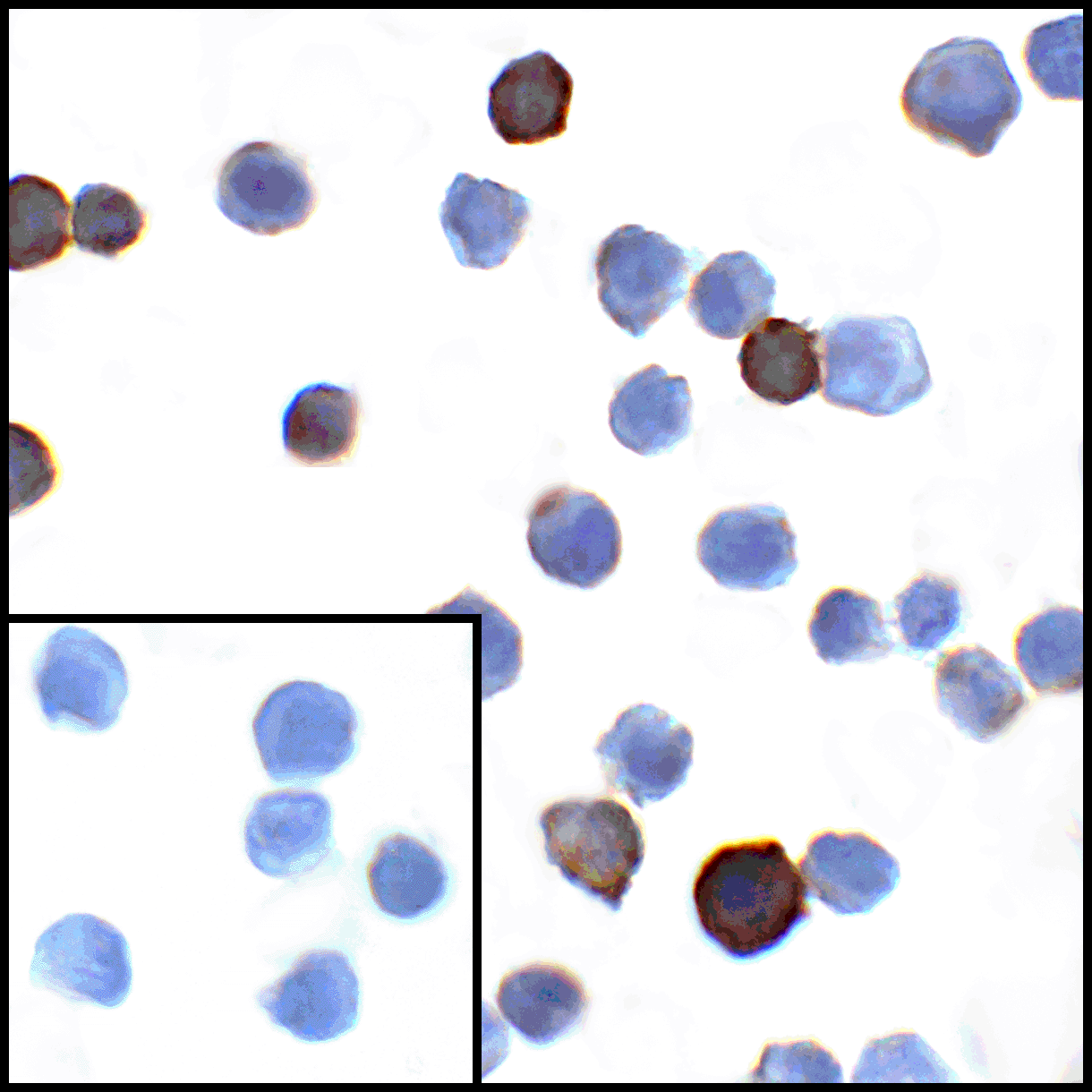
Immunocytochemistry of PD-L1 in transfected HEK293 cells with PD-L1 antibody at 1 &956,g/mL. Lower left: Immunocytochemistry in transfected HEK293 cells with control mouse IgG antibody at 1 &956,g/mL.
Immunofluorescence of PD-L1 in transfected HEK293 cells with PD-L1 antibody at 2 &956,g/mL. Red: PDL1 Antibody [1D7] (RF16038) Blue: DAPI staining
Immunofluorescence of PD-L1 in human stomach carcinoma tissue with PD-L1 antibody at 2 &956,g/mL. Red: PDL1 Antibody [1D7] (RF16038) Blue: DAPI staining
Immunofluorescence of PD-L1 in human tonsil tissue with PD-L1 antibody at 2 &956,g/mL. Red: PDL1 Antibody [1D7] (RF16038) Blue: DAPI staining
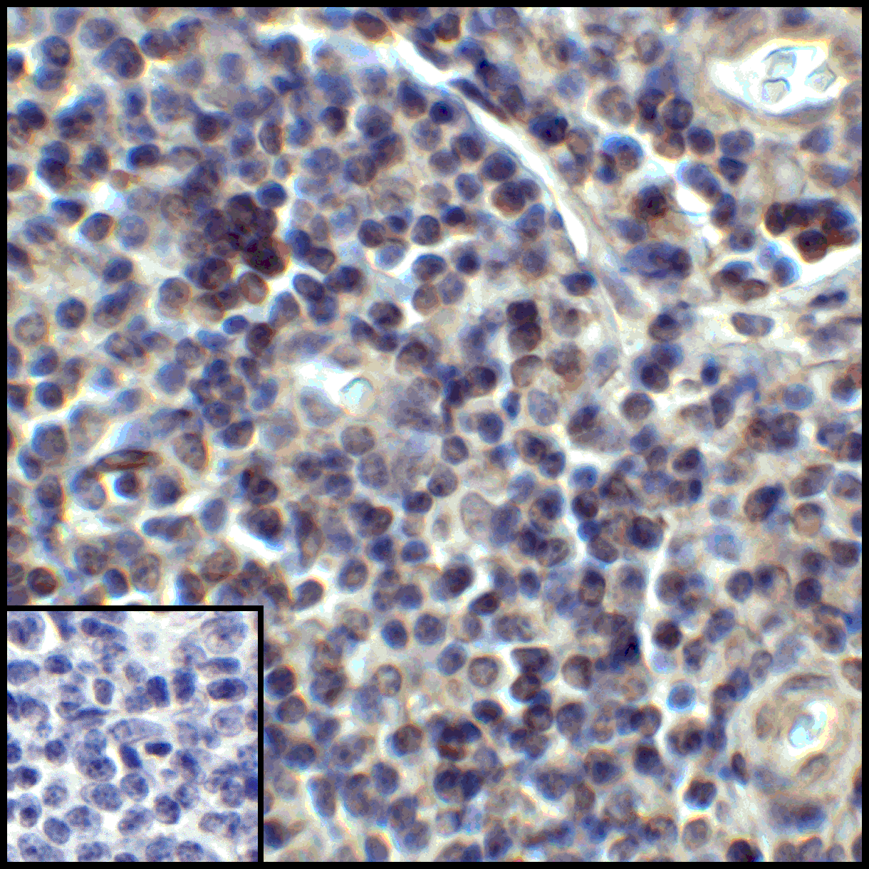
Immunohistochemistry of PD-L1 in human stomach carcinoma tissue with PD-L1 antibody at 5 &956,g/mL.
Immunohistochemistry of PD-L1 in human tonsil tissue with PD-L1 antibody at 5 &956,g/mL.
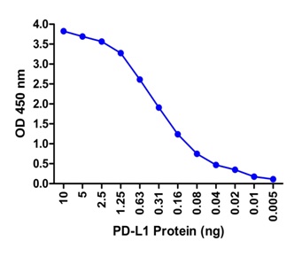
A sandwich ELISA was performed using the anti-PD-L1 mAb RF16038 (5 &956,g/ml) as the capture antibody. Biotin-labeled anti-PD-L1 mAb RF16032-biotin (1 &956,g/ml) and streptavidin-HRP (0.1 &956,g/ml) were used for detection. Detection range is from 10 ng to 20 pg.
Western blot analysis of PD-L1 in overexpressing HEK293 cells PD-L1 antibody at 0.25 and 0.5 &956,g/ml
* VAT and and shipping costs not included. Errors and price changes excepted