
LRPPRC Rabbit pAb, Unconjugated, Polyclonal
Artikelnummer:
ABB-A3365
- Bilder (8)
| Artikelname: | LRPPRC Rabbit pAb, Unconjugated, Polyclonal |
| Artikelnummer: | ABB-A3365 |
| Hersteller Artikelnummer: | A3365 |
| Alternativnummer: | ABB-A3365-100UL,ABB-A3365-500UL,ABB-A3365-20UL,ABB-A3365-1000UL |
| Hersteller: | ABclonal |
| Wirt: | Rabbit |
| Kategorie: | Antikörper |
| Applikation: | ELISA, IF, IHC-P, WB |
| Spezies Reaktivität: | Human |
| Immunogen: | Recombinant protein (or fragment).This information is considered to be commercially sensitive. |
| Konjugation: | Unconjugated |
| Alternative Synonym: | LSFC, GP130, LRP130, MC4DN5, CLONE-23970, LRPPRC |
| This gene encodes a leucine-rich protein that has multiple pentatricopeptide repeats (PPR). The precise role of this protein is unknown but studies suggest it may play a role in cytoskeletal organization, vesicular transport, or in transcriptional regulation of both nuclear and mitochondrial genes. The protein localizes primarily to mitochondria and is predicted to have an N-terminal mitochondrial targeting sequence. Mutations in this gene are associated with the French-Canadian type of Leigh syndrome. |
| Klonalität: | Polyclonal |
| Molekulargewicht: | 158kDa |
| NCBI: | 10128 |
| UniProt: | P42704 |
| Reinheit: | Affinity purification |
| Sequenz: | IACRLNQKKGAYDIFLNAKEQNIVFNAETYSNLIKLLMSEDYFTQAMEVKAFAETHIKGFTLNDAANSRLIITQVRRDYLKEAVTTLKTVLDQQQTPSRLAVTRVIQALAMKGDVENIEVVQKMLNGLEDSIGLSKMVFINNIALAQIKNNNIDAAIENIENMLTSENKVIEPQYFGLAYLFRKVIEEQLEPAVEKISIMAERLANQFAIYKPVTDFFLQLVDAGKVDDARALLQRCGAIAEQTPILLLFLLRNS |
| Target-Kategorie: | LRPPRC |
| Antibody Type: | Primary Antibody |
| Application Verdünnung: | WB,1:1000 - 1:5000|IHC-P,1:50 - 1:200|IF/ICC,1:100 - 1:200|ELISA,Recommended starting concentration is 1 µg/mL. Please optimize the concentration based on your specific assay requirements. |
| Anwendungsbeschreibung: | Cross-Reactivity: Human,Mouse,Rat. ResearchArea: Epigenetics Nuclear Signaling,RNA Binding,Endocrine Metabolism,Mitochondrial metabolism,Neuroscience,Neurodegenerative Diseases. Shipping: Ice Bag |
|
|
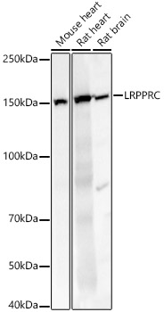
Western blot analysis of various lysates, using LRPPRC Rabbit pAb (A3365) at 1:2000 dilution. Secondary antibody: HRP-conjugated Goat anti-Rabbit IgG (H+L) (AS014) at 1:10000 dilution. Lysates/proteins: 25µg per lane. Blocking buffer: 3% nonfat dry milk in TBST. Detection: ECL Basic Kit (RM00020). Exposure time: 10s. |
|
|
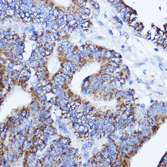
Immunohistochemistry analysis of paraffin-embedded Human breast cancer using LRPPRC Rabbit pAb (A3365) at dilution of 1:200 (40x lens). Microwave antigen retrieval performed with 0.01M PBS Buffer (pH 7.2) prior to IHC staining. |
|
|
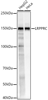
Western blot analysis of various lysates, using LRPPRC Rabbit pAb (A3365) at 1:2000 dilution. Secondary antibody: HRP-conjugated Goat anti-Rabbit IgG (H+L) (AS014) at 1:10000 dilution. Lysates/proteins: 25µg per lane. Blocking buffer: 3% nonfat dry milk in TBST. Detection: ECL Basic Kit (RM00020). Exposure time: 0.5s. |
|
|
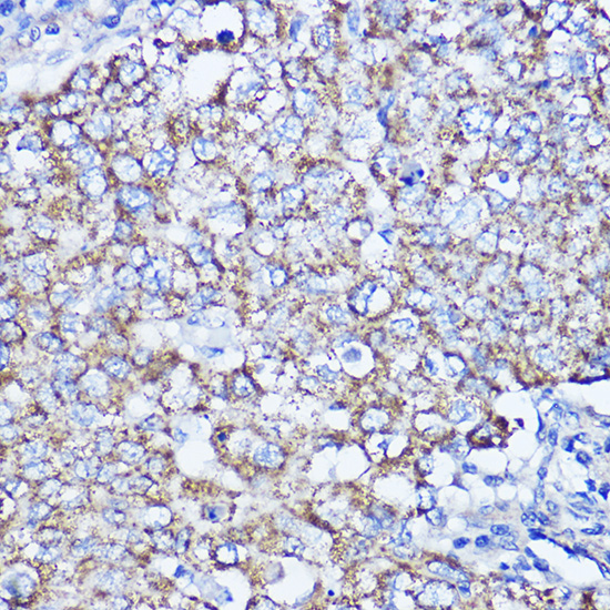
Immunohistochemistry analysis of paraffin-embedded Human colon carcinoma using LRPPRC Rabbit pAb (A3365) at dilution of 1:200 (40x lens). Microwave antigen retrieval performed with 0.01M PBS Buffer (pH 7.2) prior to IHC staining. |
|
|
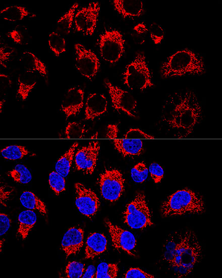
Confocal immunofluorescence analysis of Hela cells using LRPPRC Rabbit pAb (A3365) at dilution of 1:400. Blue: DAPI for nuclear staining. |
|
|
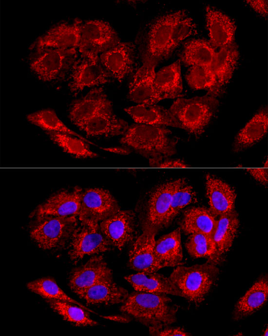
Immunofluorescence analysis of A-549 cells using LRPPRC Rabbit pAb (A3365) at dilution of 1:100 (40x lens). Secondary antibody: Cy3-conjugated Goat anti-Rabbit IgG (H+L) (AS007) at 1:500 dilution. Blue: DAPI for nuclear staining. |
|
|
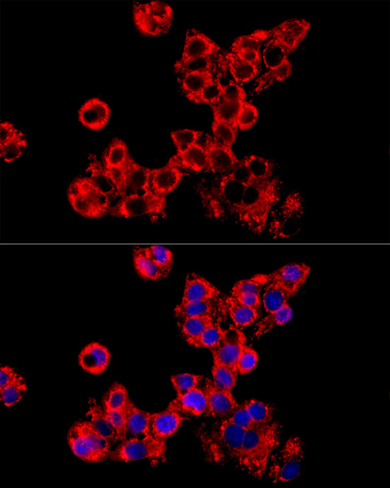
Immunofluorescence analysis of HepG2 cells using LRPPRC Rabbit pAb (A3365) at dilution of 1:100 (40x lens). Secondary antibody: Cy3-conjugated Goat anti-Rabbit IgG (H+L) (AS007) at 1:500 dilution. Blue: DAPI for nuclear staining. |
|
|
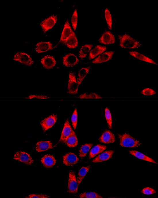
Immunofluorescence analysis of NIH/3T3 cells using LRPPRC Rabbit pAb (A3365) at dilution of 1:100 (40x lens). Secondary antibody: Cy3-conjugated Goat anti-Rabbit IgG (H+L) (AS007) at 1:500 dilution. Blue: DAPI for nuclear staining. |
Produktgarantie und fachkundiger Support
