
HSP20/HSPB6 Rabbit mAb, Unconjugated, Monoclonal
Artikelnummer:
ABB-A9091
- Bilder (9)

| Artikelname: | HSP20/HSPB6 Rabbit mAb, Unconjugated, Monoclonal |
| Artikelnummer: | ABB-A9091 |
| Hersteller Artikelnummer: | A9091 |
| Alternativnummer: | ABB-A9091-100UL,ABB-A9091-20UL |
| Hersteller: | ABclonal |
| Wirt: | Rabbit |
| Kategorie: | Antikörper |
| Applikation: | ELISA, IF, IHC-P, WB |
| Spezies Reaktivität: | Human |
| Immunogen: | Synthetic peptide. This information is considered to be commercially sensitive. |
| Konjugation: | Unconjugated |
| Alternative Synonym: | HEL55, Hsp20, PPP1R91, HSP20/HSPB6 |
| This locus encodes a heat shock protein. The encoded protein likely plays a role in smooth muscle relaxation. |
| Application Verdünnung: | WB,1:500 - 1:1000|IF-P,1:50 - 1:200|IHC-P,1:50 - 1:200|ELISA,Recommended starting concentration is 1 µg/mL. Please optimize the concentration based on your specific assay requirements. |
| Anwendungsbeschreibung: | Cross-Reactivity: Human,Mouse,Rat. ResearchArea: Apoptosis,Signal Transduction ,Protein Trafficking ,Chaperones ,Heat Shock Proteins,Cardiovascular, Atherosclerosis ,Ischemia / Reperfusion,Cardiovascular ,Heart Apoptosis. Shipping: Ice Bag |
|
|
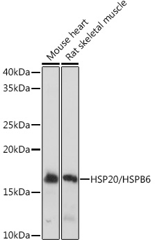
Western blot analysis of various lysates using HSP20/HSPB6 Rabbit mAb (A9091) at 1:1000 dilution. Secondary antibody: HRP-conjugated Goat anti-Rabbit IgG (H+L) (AS014) at 1:10000 dilution. Lysates/proteins: 25µg per lane. Blocking buffer: 3% nonfat dry milk in TBST. Detection: ECL Basic Kit (RM00020). Exposure time: 1s. |
|
|
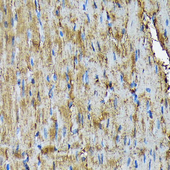
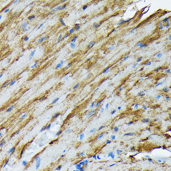
Immunohistochemistry analysis of paraffin-embeddedMouse heart tissue usingHSP20/HSPB6 Rabbit mAb(A9091) at a dilution of 1:200 (40x lens).High pressure antigen retrieval was performed with 0.01 M Tris-EDTA buffer (pH 9.0) prior to IHC staining. |
|
|
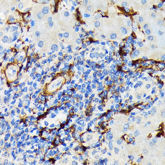
Immunohistochemistry analysis of paraffin-embeddedHuman colon carcinoma tissue usingHSP20/HSPB6 Rabbit mAb(A9091) at a dilution of 1:200 (40x lens).High pressure antigen retrieval was performed with 0.01 M Tris-EDTA buffer (pH 9.0) prior to IHC staining. |
|
|
Immunohistochemistry analysis of paraffin-embeddedHuman liver cancer tissue usingHSP20/HSPB6 Rabbit mAb(A9091) at a dilution of 1:200 (40x lens).High pressure antigen retrieval was performed with 0.01 M Tris-EDTA buffer (pH 9.0) prior to IHC staining. |
|
|
Immunohistochemistry analysis of paraffin-embeddedRat colon tissue usingHSP20/HSPB6 Rabbit mAb(A9091) at a dilution of 1:200 (40x lens).High pressure antigen retrieval was performed with 0.01 M Tris-EDTA buffer (pH 9.0) prior to IHC staining. |
|
|
Immunohistochemistry analysis of paraffin-embeddedHuman lung tissue usingHSP20/HSPB6 Rabbit mAb(A9091) at a dilution of 1:200 (40x lens).High pressure antigen retrieval was performed with 0.01 M Tris-EDTA buffer (pH 9.0) prior to IHC staining. |
|
|
Immunohistochemistry analysis of paraffin-embeddedRat heart tissue usingHSP20/HSPB6 Rabbit mAb(A9091) at a dilution of 1:200 (40x lens).High pressure antigen retrieval was performed with 0.01 M Tris-EDTA buffer (pH 9.0) prior to IHC staining. |
|
|
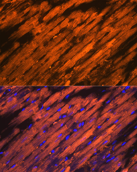
Confocal imaging ofparaffin-embedded Rat heart tissue usingHSP20/HSPB6 Rabbit mAb (A9091, dilution 1:200) followed by a further incubation with Cy3 Goat Anti-Rabbit IgG (H+L) (AS007, dilution 1:500) (Red). DAPI was used for nuclear staining (Blue). Objective: 40x. Perform high pressure antigen retrieval with 0.01 M citrate buffer (pH 6.0) prior to IF staining. |
|
|
Confocal imaging ofparaffin-embedded Mouse heart tissue usingHSP20/HSPB6 Rabbit mAb (A9091, dilution 1:200) followed by a further incubation with Cy3 Goat Anti-Rabbit IgG (H+L) (AS007, dilution 1:500) (Red). DAPI was used for nuclear staining (Blue). Objective: 40x. Perform high pressure antigen retrieval with 0.01 M citrate buffer (pH 6.0) prior to IF staining. |
Produktgarantie und fachkundiger Support
