

Anti-SAMHD1 Antibody, Rabbit, Polyclonal
Artikelnummer:
BOB-A00592-1
- Bilder (9)
| Artikelname: | Anti-SAMHD1 Antibody, Rabbit, Polyclonal |
| Artikelnummer: | BOB-A00592-1 |
| Hersteller Artikelnummer: | A00592-1 |
| Alternativnummer: | BOB-A00592-1-0.1MG |
| Hersteller: | Boster Bio |
| Wirt: | Rabbit |
| Kategorie: | Antikörper |
| Applikation: | ELISA, IF, IHC, WB |
| Spezies Reaktivität: | Human, Mouse |
| Immunogen: | Anti-SAMHD1 antibody was raised against a peptide corresponding to 18 amino acids near the carboxy terminus of human SAMHD1. |
| Alternative Synonym: | SAM domain and HD domain 1, DCIP, CHBL2, HDDC1, MOP-5, SBBI88 |
| Boster Bio Anti-SAMHD1 Antibody (Catalog A00592-1). Tested in ELISA, WB, IHC, IF applications. This antibody reacts with Human, Mouse. |
| Klonalität: | Polyclonal |
| Konzentration: | 1 mg/mL |
| Molekulargewicht: | 72201 MW |
| UniProt: | Q9Y3Z3 |
| Puffer: | SAMHD1 antibody is supplied in PBS containing 0.02% sodium azide. |
| Formulierung: | Liquid |
| Application Verdünnung: | WB: 1 µg/mL, IHC: 5 µg/mL, IF: 20 µg/mL.Antibody validated: Western Blot, Immunohistochemistry, Immunofluorescence in human samples. All other applications and species not yet tested. Optimal dilutions for each application should be determined by the rese |
|
|
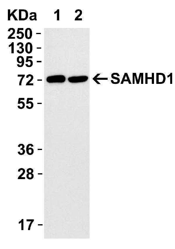
Overexpression Validation in 293T Transfected CellsLoading: 15 &956,g of lysates per lane.Antibodies: SAMHD1 A00592-1 (0.1 &956,g/mL), (1h incubation at RT in 5% NFDM/TBST.Secondary: Goat anti-rabbit IgG HRP conjugate at 1:10000 dilution.293 cells were transfected with (1) wild-type SAMHD1 or (2) SAMHD1 (mutation T592A). |
|
|
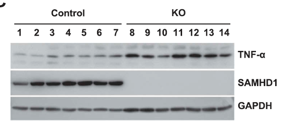
KO Validation of SAMHD1 in xenograft mice (Kodigepalli et al., 2018) THP-1 control and SAMHD1 KO (THP-1/KO) cells were injected into NSG (non-obese diabetic/severe combinedimmune deficient-gamma) mice. Protein expression levels of SAMHD1 were examined by Western blot with anti-SAMHD1 antibodies (A00592-1) and SAMHD1 was not detected in THP-1/KO cells. |
|
|
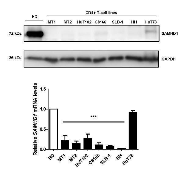
Overexpression Validation of SAMHD1 in CD4+ T-cells from a healthy donor and transformed CD4+ T-cell lines (Kohnken et al., 2017) MT1, MT2, SLB-1, and C8166 were from leukemia patients and HH, HuT78, and HuT102 were from cutaneous T-cell lymphoma (CTCL) patients. SAMHD1 protein expression detected by anti-SAMHD1 antibodies (A00592-1) was significantly increased in normal CD4+ T-cells as compared to leukemia- and CTCL- derived CD4+ T-cell lines. This blot is representative from four independent experiments with four healthy donors. |
|
|
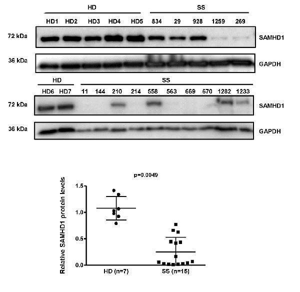
Overexpression of SAMHD1 in CD4+ T-cells from Healthy Donors and Sezary Syndrome (SS) patients(Kohnken et al., 2017)SAMHD1 protein expression detected by anti-SAMHD1 antibodies (A00592-1) was significantly reduced in CD+ T-cells from 15 SS patients as compared to those from 7 healthy donors. |
|
|
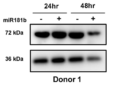
Regulated Expression Validation of SAMHD1 in CD4+ T-cells from a healthy donor (Kohnken et al., 2017)SAMHD1 protein expression detected by anti-SAMHD1 antibodies (A00592-1) was significantly decreased by about 40% relative to control cells at 48hr post-nucleofection with miR-181b. |
|
|
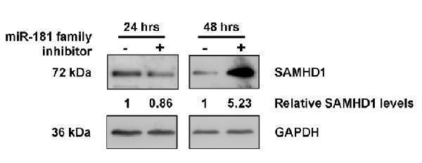
Regulated Expression Validation of SAMHD1 in MT2 CD4+ T-cells from leukemia patients(Kohnken et al., 2017)SAMHD1 protein expression detected by anti-SAMHD1 antibodies (A00592-1) was significantly increased by 5-fold at 48hr post-nucleofection with miR-181family inhibitor treatment. |
|
|
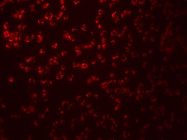
Immunofluorescence Validation of SAMHD1 in Human Daudi cellsImmunofluorescent analysis of 4% paraformaldehyde-fixed Human Daudi Cells labeling SAMHD1 with A00592-1 at 20 &956,g/mL, followed by goat anti-rabbit IgG secondary antibody at 1/500 dilution (red). |
|
|
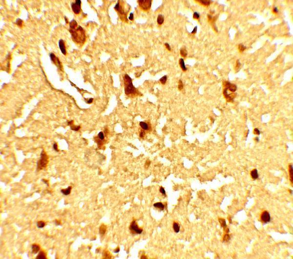
Immunohistochemistry Validation of SAMHD1 in Human Brain Tissue Immunohistochemical analysis of paraffin-embedded Human Brain Tissue using anti- SAMHD1 antibody (A00592-1) at 5 &956,g/ml. Tissue was fixed with formaldehyde and blocked with 10% serum for 1 h at RT, antigen retrieval was by heat mediation with a citrate buffer (pH6). Samples were incubated with primary antibody overnight at 4 &730,C. A goat anti-rabbit IgG H&L (HRP) at 1/250 was used as secondary. Counter stained with Hematoxylin. |
|
|
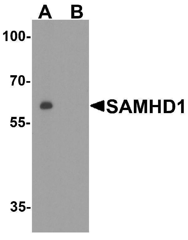
Western Blot Validation in Human Daudi Cell LinesLoading: 15 &956,g of lysates per lane.Antibodies: SAMHD1 A00592-1, 1 &956,g/mL, in (A: the absence and B: the presence of blocking peptide), (1h incubation at RT in 5% NFDM/TBST.Secondary: Goat anti-rabbit IgG HRP conjugate at 1:10000 dilution. |
Produktgarantie und fachkundiger Support
