
Hemoglobin subunit alpha (HBA1) Rabbit mAb, Unconjugated, Monoclonal
Catalog Number:
ABB-A9293
- Images (9)
| Article Name: | Hemoglobin subunit alpha (HBA1) Rabbit mAb, Unconjugated, Monoclonal |
| Biozol Catalog Number: | ABB-A9293 |
| Supplier Catalog Number: | A9293 |
| Alternative Catalog Number: | ABB-A9293-100UL,ABB-A9293-20UL,ABB-A9293-1000UL,ABB-A9293-500UL |
| Manufacturer: | ABclonal |
| Host: | Rabbit |
| Category: | Antikörper |
| Application: | ELISA, IF, IHC-P, WB |
| Species Reactivity: | Human |
| Immunogen: | Synthetic peptide. This information is considered to be commercially sensitive. |
| Conjugation: | Unconjugated |
| Alternative Names: | HBH, ECYT7, HBA-T3, METHBA, Hemoglobin subunit alpha (HBA1) |
| The human alpha globin gene cluster located on chromosome 16 spans about 30 kb and includes seven loci: 5- zeta - pseudozeta - mu - pseudoalpha-1 - alpha-2 - alpha-1 - theta - 3. The alpha-2 (HBA2) and alpha-1 (HBA1) coding sequences are identical. These genes differ slightly over the 5 untranslated regions and the introns, but they differ significantly over the 3 untranslated regions. Two alpha chains plus two beta chains constitute HbA, which in normal adult life comprises about 97% of the total hemoglobin, alpha chains combine with delta chains to constitute HbA-2, which with HbF (fetal hemoglobin) makes up the remaining 3% of adult hemoglobin. Alpha thalassemias result from deletions of each of the alpha genes as well as deletions of both HBA2 and HBA1, some nondeletion alpha thalassemias have also been reported. |
| Application Dilute: | WB,1:1000 - 1:6000|IF-P,1:100 - 1:400|IHC-P,1:200 - 1:2000|ELISA,Recommended starting concentration is 1 µg/mL. Please optimize the concentration based on your specific assay requirements. |
| Application Notes: | Cross-Reactivity: Human,Mouse,Rat. ResearchArea: Cardiovascular,Blood,Blood Cell Antigens,RBC Antigens. Shipping: Ice Bag |
|
|
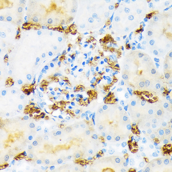
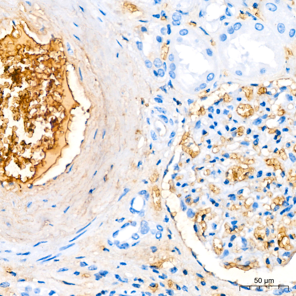
Immunohistochemistry analysis of paraffin-embedded Human kidney tissue using Hemoglobin subunit alpha (HBA1) Rabbit mAb (A9293) at a dilution of 1:200 (40x lens). High pressure antigen retrieval performed with 0.01M Citrate Buffer (pH 6.0) prior to IHC staining. |
|
|
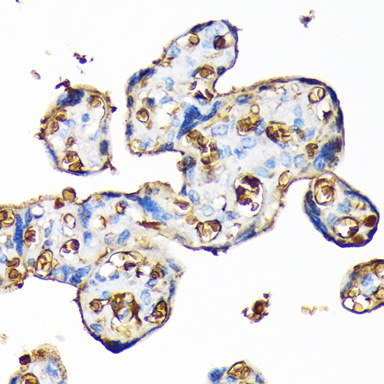
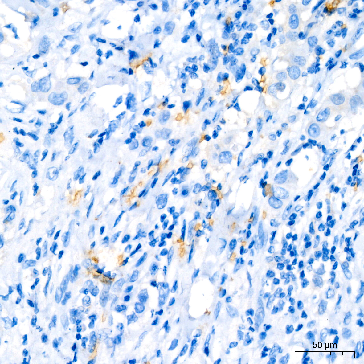
Immunohistochemistry analysis of paraffin-embedded Human breast cancer tissue using Hemoglobin subunit alpha (HBA1) Rabbit mAb (A9293) at a dilution of 1:200 (40x lens). High pressure antigen retrieval performed with 0.01M Citrate Buffer (pH 6.0) prior to IHC staining. |
|
|
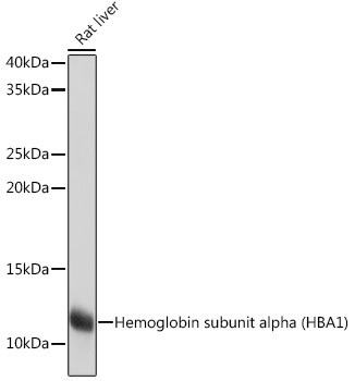
Western blot analysis of lysates from Rat liver using Hemoglobin subunit alpha (HBA1) Rabbit mAb (A9293) at 1:1000 dilution incubated overnight at 4°C. Secondary antibody: HRP-conjugated Goat anti-Rabbit IgG (H+L) (AS014) at 1:10000 dilution. Lysates/proteins: 25 µg per lane. Blocking buffer: 3% nonfat dry milk in TBST. Detection: ECL Basic Kit (RM00020). Exposure time: 1s. |
|
|
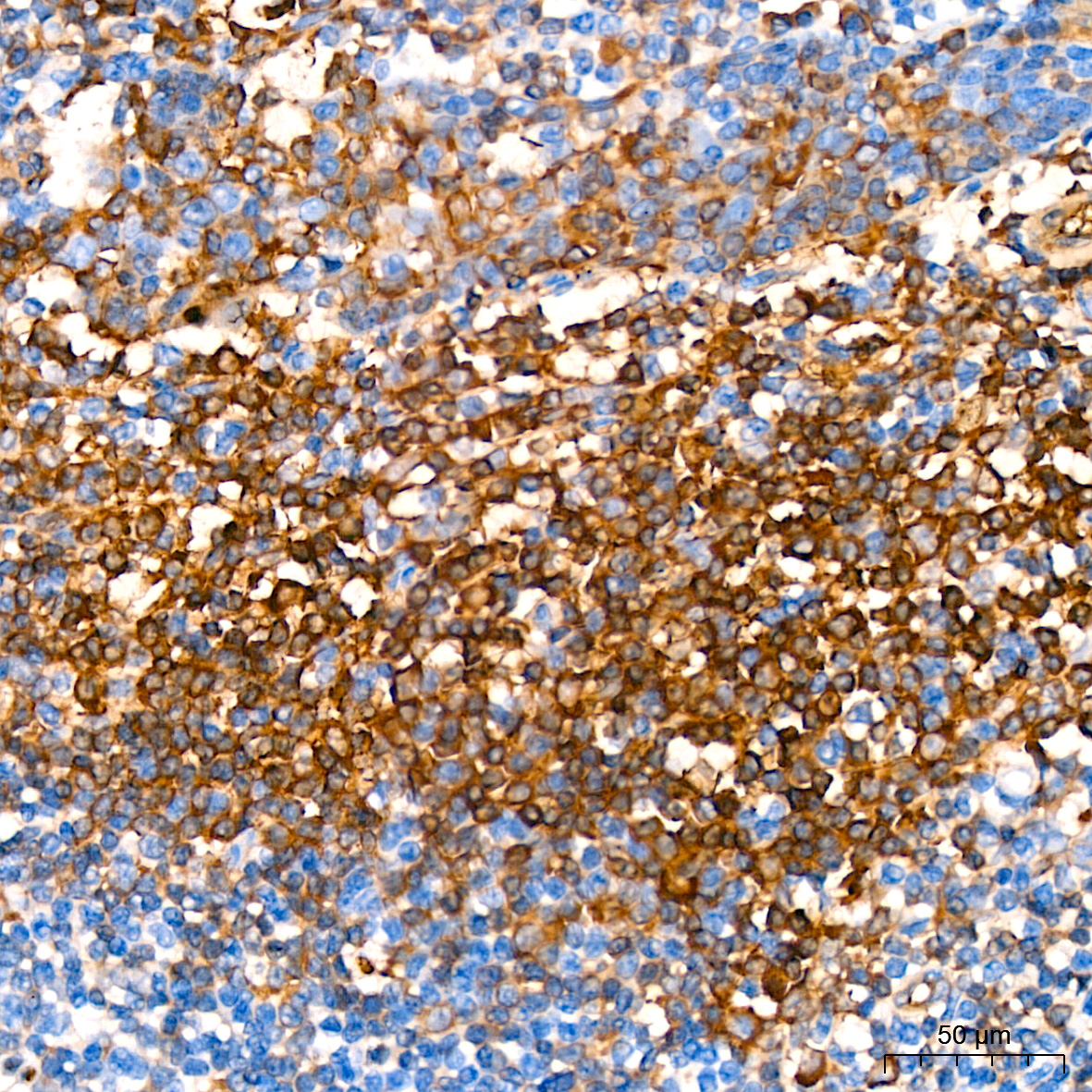
Immunohistochemistry analysis of paraffin-embedded Human tonsil tissue using Hemoglobin subunit alpha (HBA1) Rabbit mAb (A9293) at a dilution of 1:200 (40x lens). High pressure antigen retrieval performed with 0.01M Citrate Buffer (pH 6.0) prior to IHC staining. |
|
|
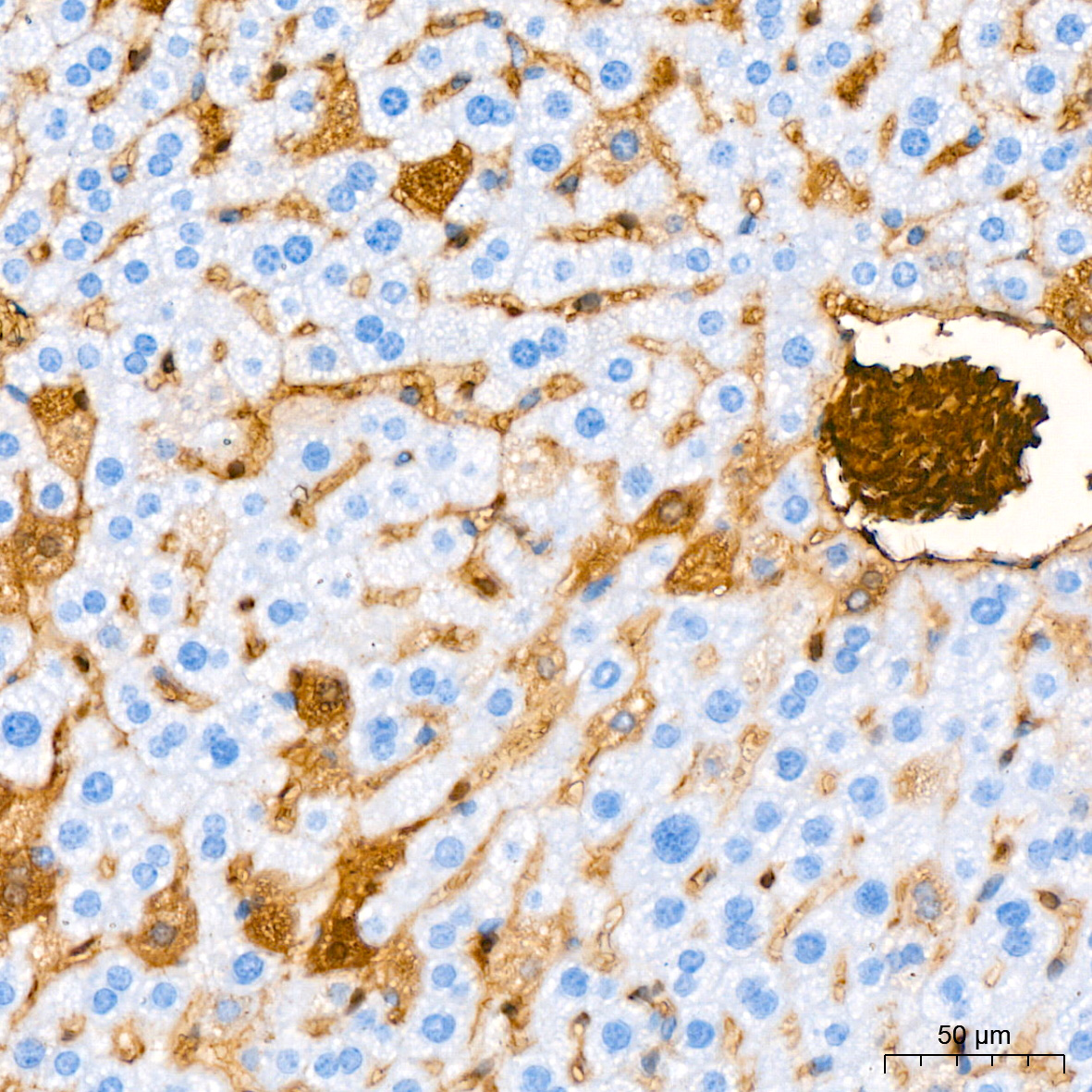
Immunohistochemistry analysis of paraffin-embedded Mouse liver tissue using Hemoglobin subunit alpha (HBA1) Rabbit mAb (A9293) at a dilution of 1:200 (40x lens). High pressure antigen retrieval performed with 0.01M Citrate Buffer (pH 6.0) prior to IHC staining. |
|
|
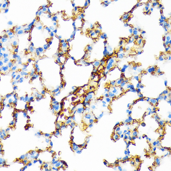
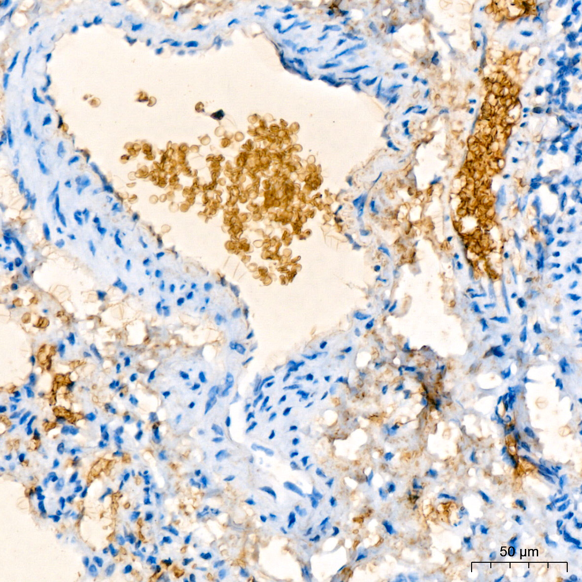
Immunohistochemistry analysis of paraffin-embedded Rat lung tissue using Hemoglobin subunit alpha (HBA1) Rabbit mAb (A9293) at a dilution of 1:200 (40x lens). High pressure antigen retrieval performed with 0.01M Citrate Buffer (pH 6.0) prior to IHC staining. |
|
|
Immunofluorescence analysis of paraffin-embedded rat spleen using Hemoglobin subunit alpha (HBA1) Rabbit mAb (A9293) at dilution of 1:100 (40x lens). Secondary antibody: Cy3-conjugated Goat anti-Rabbit IgG (H+L) (AS007) at 1:500 dilution. Blue: DAPI for nuclear staining. |
|
|
Immunofluorescence analysis of paraffin-embedded human spleen using Hemoglobin subunit alpha (HBA1) Rabbit mAb (A9293) at dilution of 1:100 (40x lens). Secondary antibody: Cy3-conjugated Goat anti-Rabbit IgG (H+L) (AS007) at 1:500 dilution. Blue: DAPI for nuclear staining. |
|
|
Immunofluorescence analysis of paraffin-embedded mouse spleen using Hemoglobin subunit alpha (HBA1) Rabbit mAb (A9293) at dilution of 1:100 (40x lens). Secondary antibody: Cy3-conjugated Goat anti-Rabbit IgG (H+L) (AS007) at 1:500 dilution. Blue: DAPI for nuclear staining. |
Product Guarantee and Expert Support
